

Forebrain > Telencephalon > Olfactory Bulb

Schematic showing the position of the olfactory bulbs in sagittal, horizontal and coronal sections through the zebrafish brain. Based on the anatomical segmentation of 3 day old zebrafish larval brain by Thomas Müller, Olaf Ronneberger, Wolfgang Driever and colleagues. For details see Ronneberger et al., Nat. Meth. 2012 and http://vibez.informatik.uni-freiburg.de
Abbreviations: Ce, cerebellar plate; D, dorsal telencephalon/pallium; E, epiphysis; EmT, eminentia thalami; Hb, habenula; Hyp, hypothalamus; MO, medulla oblongata; OB, olfactory bulb; OT, optic tectum; PG, preglomerular complex; PO, preoptic area;PrT, pretectum; PTd, posterior tuberculum dorsal part; PTh, prethalamus; PTv posterior tuberculum ventral part; Teg, tegmentum; Th, thalamus; TS, torus semicircularis; V, ventral telencephalon/subpallium; Va, valvula cerebelli.
Description
In the adult, the olfactory bulbs are paired protrusions in the anteriormost region of the telencephalon. They recieve primary sensory input from the olfactory epithelium directly via the olfactory nerve (Cranial nerve I). Cytoarchitecturally the bulb is arranged into four concentric layers.

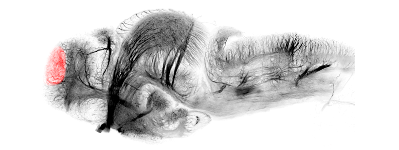
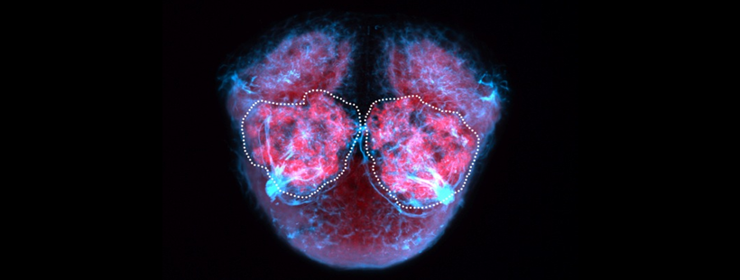
Lateral view of a 5dpf zebrafish brain labelled with SV2 and tubulin antibodies showing olfactory nerve(blue) and glomeruli (pink).

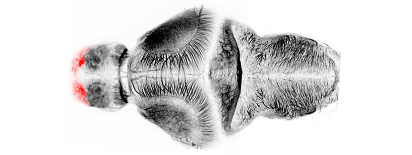
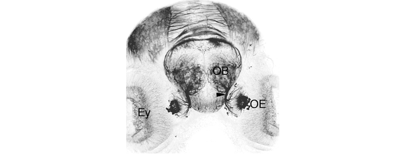
Frontal view of 4dpf zebrafish labelled with anti-tubulin showing the olfactory nerve emanating from the olfactory epithelium and entering the bulb where individual olfactory sensory neurons contact individual olfactory glomeruli.
Neuroanatomy
In the olfactory bulb there are three main cell types: mitral cells, ruffed cells and granule cells. In addition to these there is also a population of catchecholaminergic cells that are often referred to as either juxtaglomerular or periglomerular cells (Byrd and Brunjes, 1995; Fuller et al., 2005 and 2006)).
In the mature olfactory bulb, the different cell types that form the olfactory bulb are organised into four concentric layers (from external to internal layers).
1) The Primary Olfactory Fiber Layer (OFL)
This is the outermost layer of the olfactory bulb and contains axon terminals of the olfactory sensory neurons (OSNs). Each of these OSNs expresses a single olfactory receptor (OR) on its surface in the olfactory epithelium. An enormous array of odorants can be detected by the large repertoire of different ORs expressed by OSNs (Sato et al., 2007). The axons of the OSNs come together and fasciculate to form the olfactory nerve (Cranial Nerve I) that projects into the olfactory bulb. Once inside the bulb the olfactory nerve axons defasciculate and spread over the glomerular layer of the olfactory bulb to form the outer layer of primary olfactory afferents.
2) The Glomerular Layer (GL)
Primary olfactory afferents branch conspicuously and synapse onto the dendrites of the mitral cells to form a specialised spherical structure named an olfactory glomerulus. The layer containing all these olfactory glomeruli is known as the glomerular layer.
3) The Mitral or External Cellular Layer (MCL)
The Mitral or External Cellular layer is formed mainly by mitral cells, although ruffed cells can also be found in this layer (Fuller et al., 2005 and 2006). Both cell types are projection neurons and use glutamate as a neurotransmitter. In fish, individual mitral cells project several dendrites onto multiple glomeruli, whereas in mammals, single mitral cells project a primary dendrite onto a single glomerulus. Thus, a single fish mitral cell receives sensory information from several different odorant receptors (Yoshihara et al, 2001).
The apical dendrites of the mitral cells ramify and establish synaptic contact with axons of olfactory receptors to form the olfactory glomeruli. Their axons project via the lateral and medial olfactory tracts to bilaterally symmetric fields within the telencephalic lobes. These telencephalic regions are interconnected by mitral cell axonal processes that project through the anterior commissure. The fasciculated axons of the mitral cells extend posteriorly and dorsally to the habenula via the stria medularis where they cross to the contralateral side via the habenula commisure (Miyasaka, 2009). Some of these axons asymmetrically innervate the medial portion of the central neuropil domain of the right habenula. These mitral cells are labelled in the transgenic line Tg(lhx2A:GapYFP) (Miyasaka et al, 2009).
Ruffed cells are less numerous than mitral cells. Ruffed cells have short collaterals in the proximal region of their axon, which gives these cells their characteristic appearance (Fuller et al., 2005).
4) The Granule or Internal Cellular layer (GCL)
The Granule cell layer or Internal Cellular layer is mainly composed of granule cells, which act as interneurons in the olfactory bulb circuitry. These cells have apical dendrites that contact mitral and ruffed cells, while their basal dendrites contact axons originating from extrabulbar regions. They express the neurotransmitter gamma-aminobutyric acid (GABA).
A subpopulation of granule cells produce catecholamines and are tyrosine hydroxylase positive. These cells are referred to as juxtaglomerular, periglomerular or perinest cells (Byrd and Brunjes, 1995; Fuller et al., 2005 and 2006).
The adult zebrafish OB contains another subset of neuroendocrine cells that express Gonadotrophin releasing hormone(GnRH). These cells are known as terminal nerve cells. These neurons are born near to the olfactory epithelium and then migrate along the axons of the olfactory sensory neurons to populate the bulb (Whitlock et al., 2003; Palevitch et al., 2007).

Schematic of olfactory bulb cytoarchitecture
abbreviations: GC granule cell; GL glomerular layer; ICL inner cellular layer; MC mitral cell;MCL mitral cell layer; NI cranial nerve I/olfactory nerve; ONL outer layer of primary olfactory afferents; OSN olfactory sensory neuron; PC periglomerular cell.
Development
The olfactory bulbs along with the rest of the telencephalic lobes originate from the anterior neural plate.
At 1dpf and 2dpf, the prospective olfactory bulb is located in a dorsoposterior region of the telencephalon, adjacent to the diencephalon. From 2dpf to 5dpf, an anterior morphogenetic movement brings the olfactory bulb rostrally to its characteristic position as the anteriormost structure of the brain and away from the diencephalon.
By 2dpf all major neuronal types are present and have elaborated processes so that a number of glomeruli can also be seen.
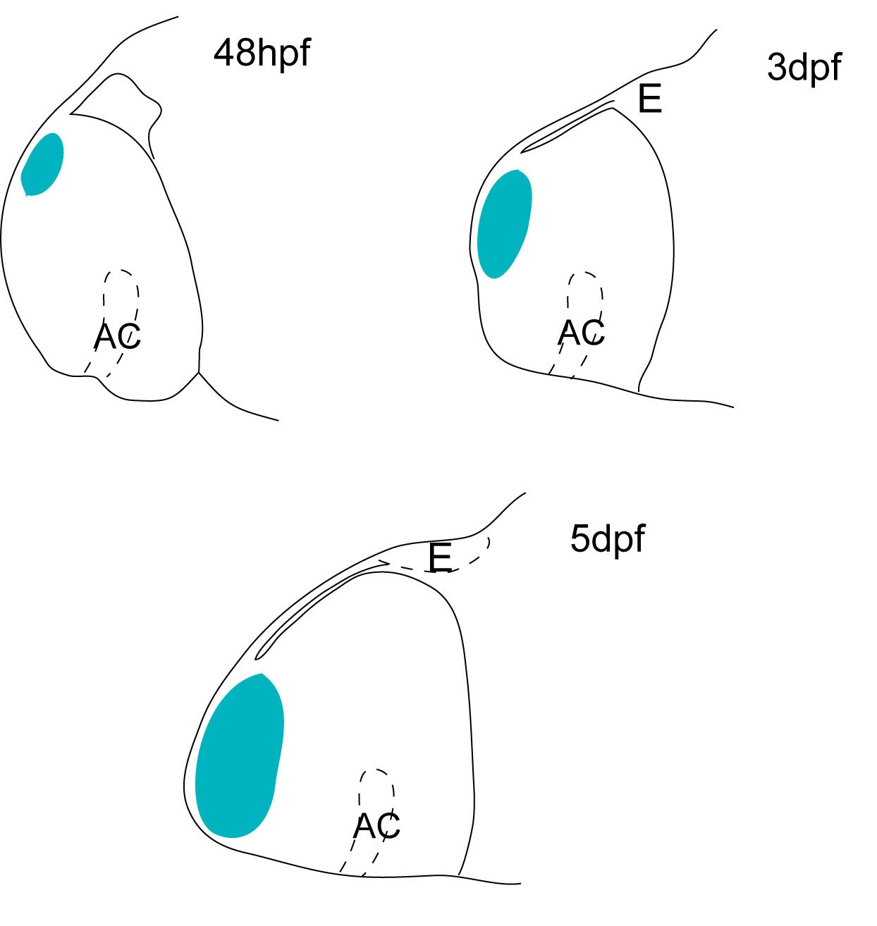
Cartoon showing the position of the olfactory bulb at three different stages of the zebrafish development (2dpf, 3dpf and 5dpf). The olfactory bulb is located in a dorso-posterior position in the telencephalon at 2dpf, but later in development (5dpf) acquires its characteristic anterior position.

Ontology
is part of: telencephalon
has parts: external cellular layer, glomerular layer, internal cellular layer, primary olfactory fiber layer
Transgenic Lines that label this brain region


Antibodies that label this brain region


Key Publications
Miyasaka, N., Morimoto, K., Tsubokawa, T., Higashijima, S., Okamoto, H., and Yoshihara, Y. (2009)
From the olfactory bulb to higher brain centers: genetic visualization of secondary olfactory pathways in zebrafish.
The Journal of neuroscience 29(15):4756-4767.
Miyasaka, N., Wanner, A.A., Li, J., Mack-Bucher, J., Genoud, C., Yoshihara, Y., and Friedrich, R.W. (2013)
Functional development of the olfactory system in zebrafish.
Mechanisms of Development. 130(6-8):336-46.
Miyasaka, N., Arganda-Carreras, I., Wakisaka, N., Masuda, M., Sümbül, U., Seung, H.S., Yoshihara, Y. (2014)
Olfactory projectome in the zebrafish forebrain revealed by genetic single-neuron labelling. Nature communications. 5:3639.
Miyasaka, N., Sato, Y., Yeo, S.Y., Hutson, L.D., Chien, C.B., Okamoto, H., and Yoshihara, Y. (2005) Robo2 is required for establishment of a precise glomerular map in the zebrafish olfactory system.
Development (Cambridge, England). 132(6):1283-1293.
Back to Forebrain