About
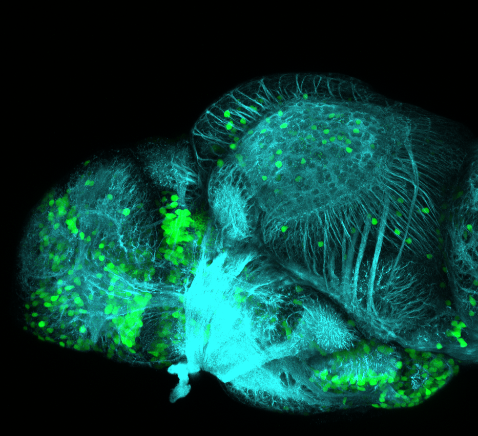
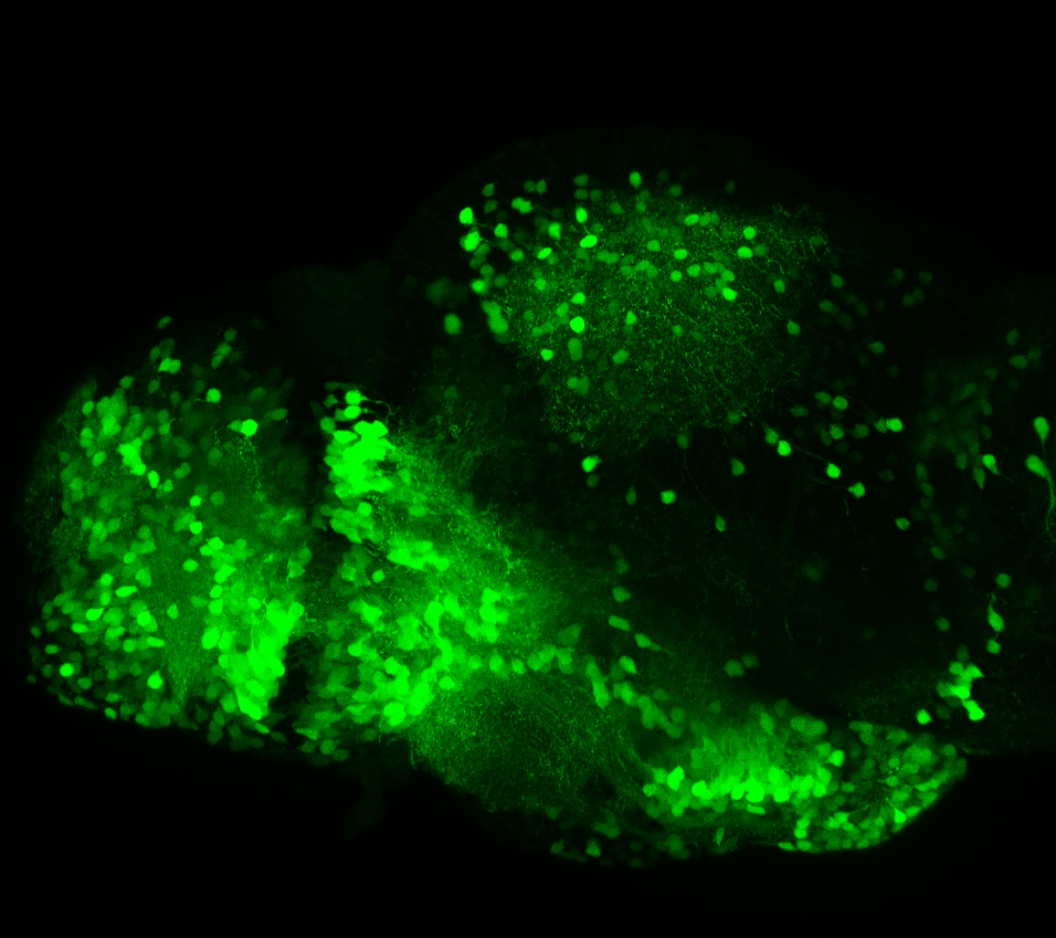
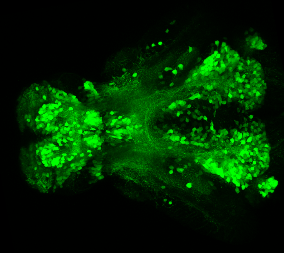
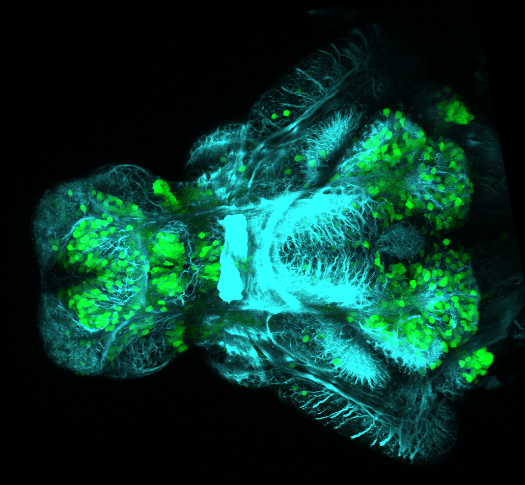
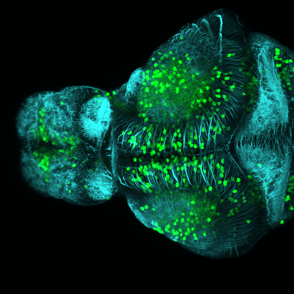
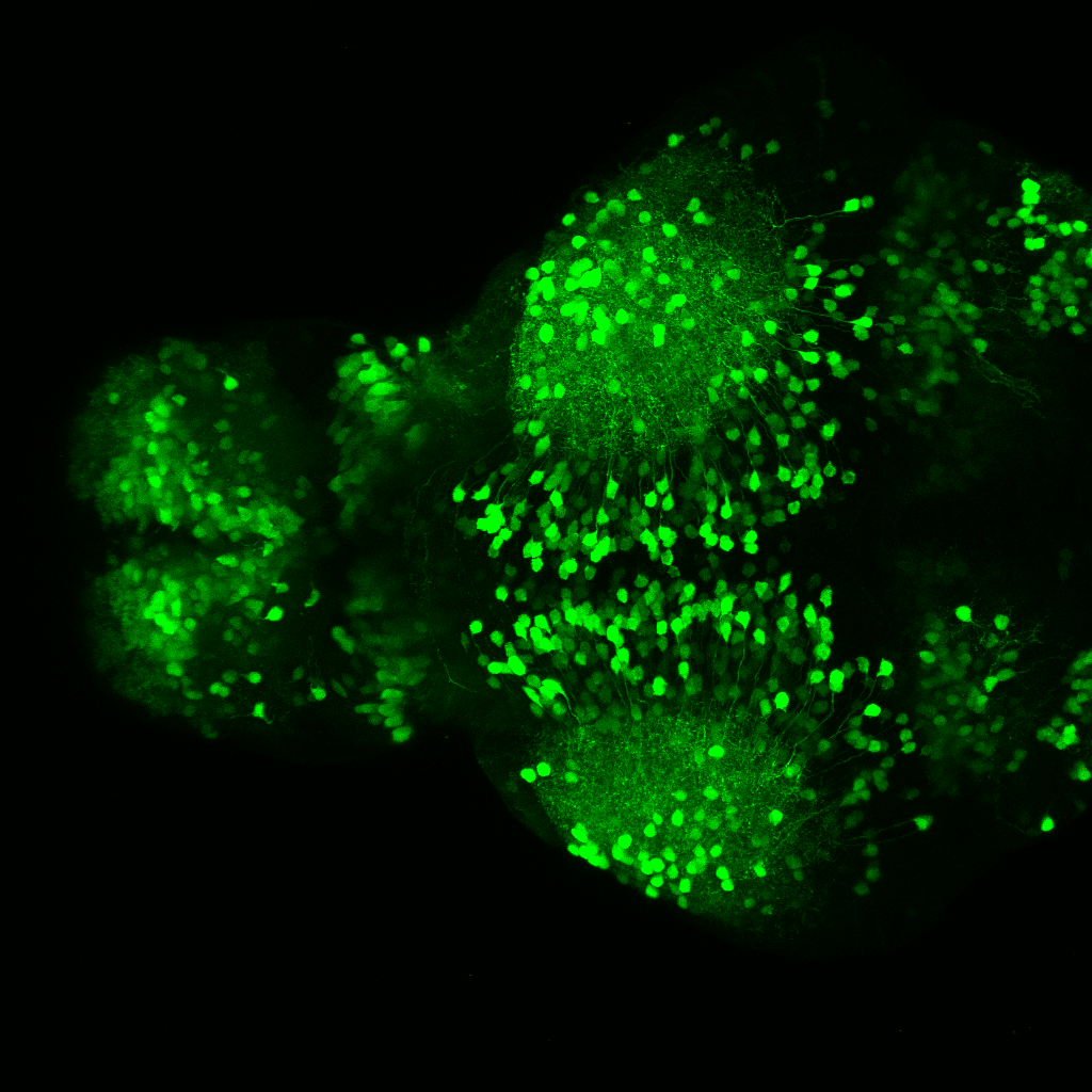
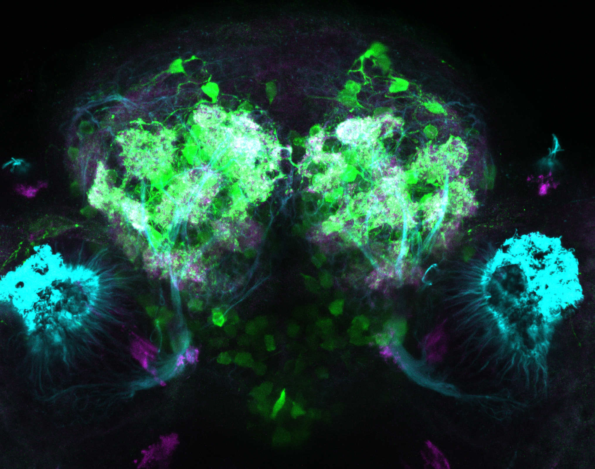
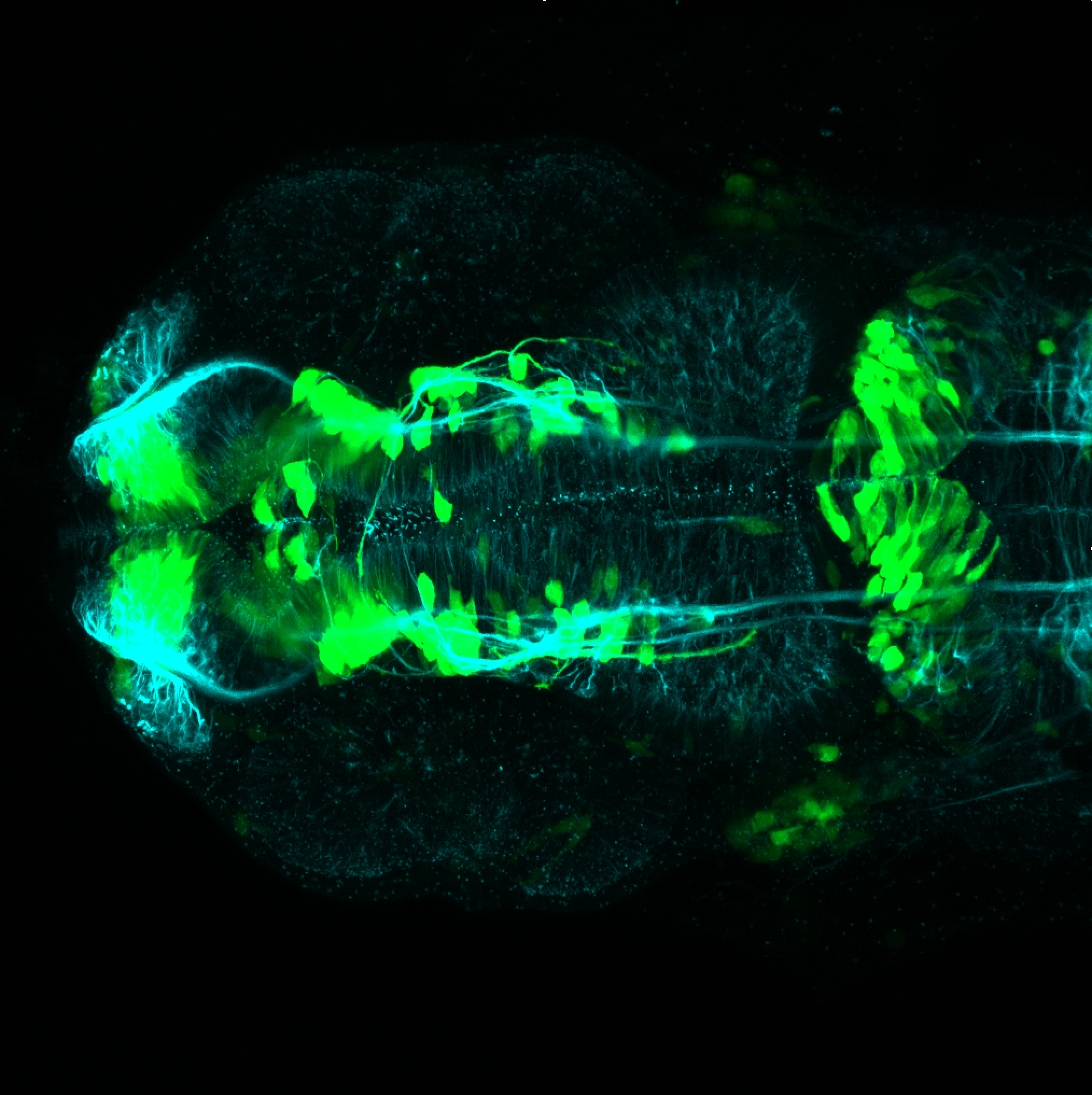
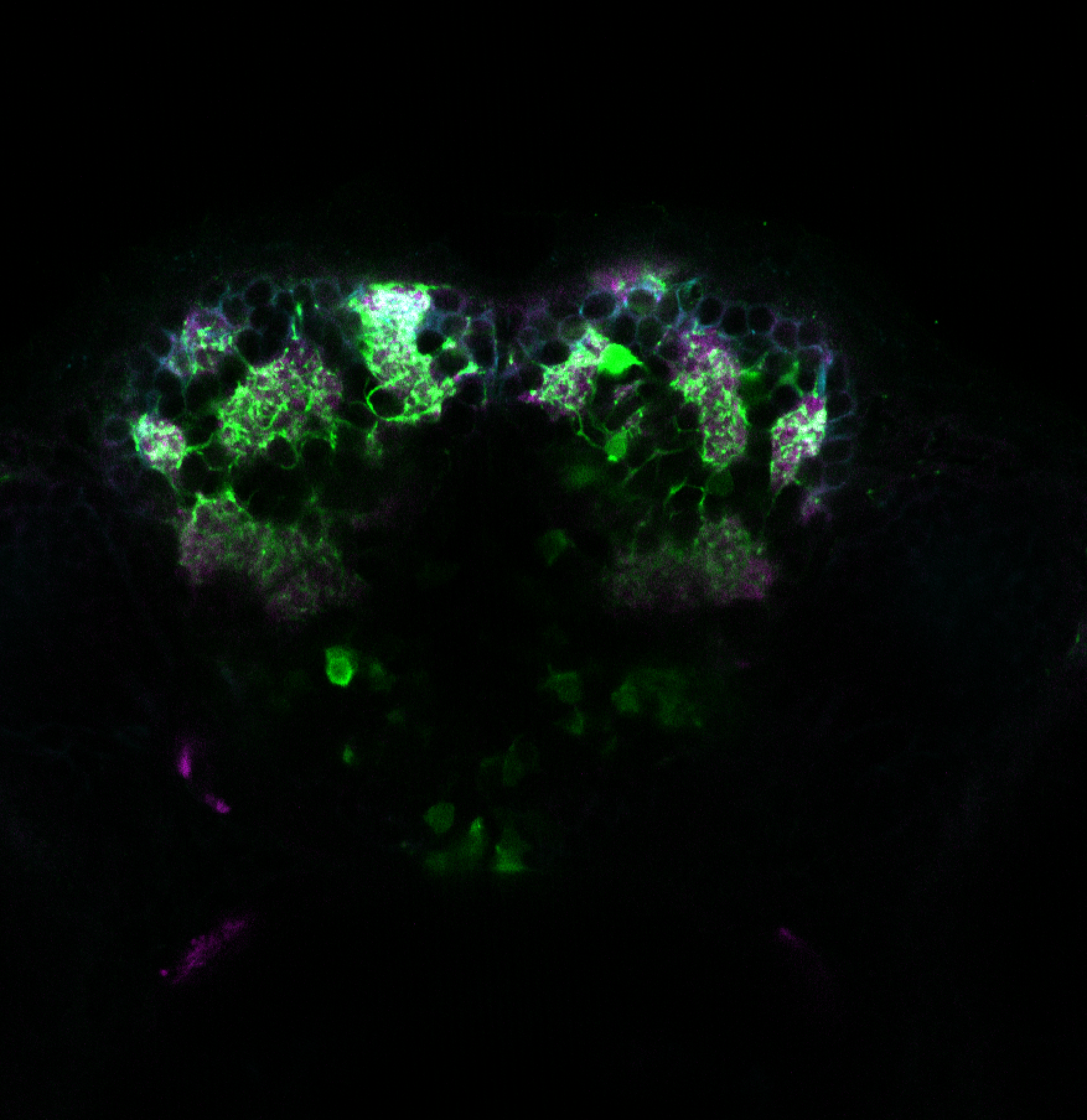
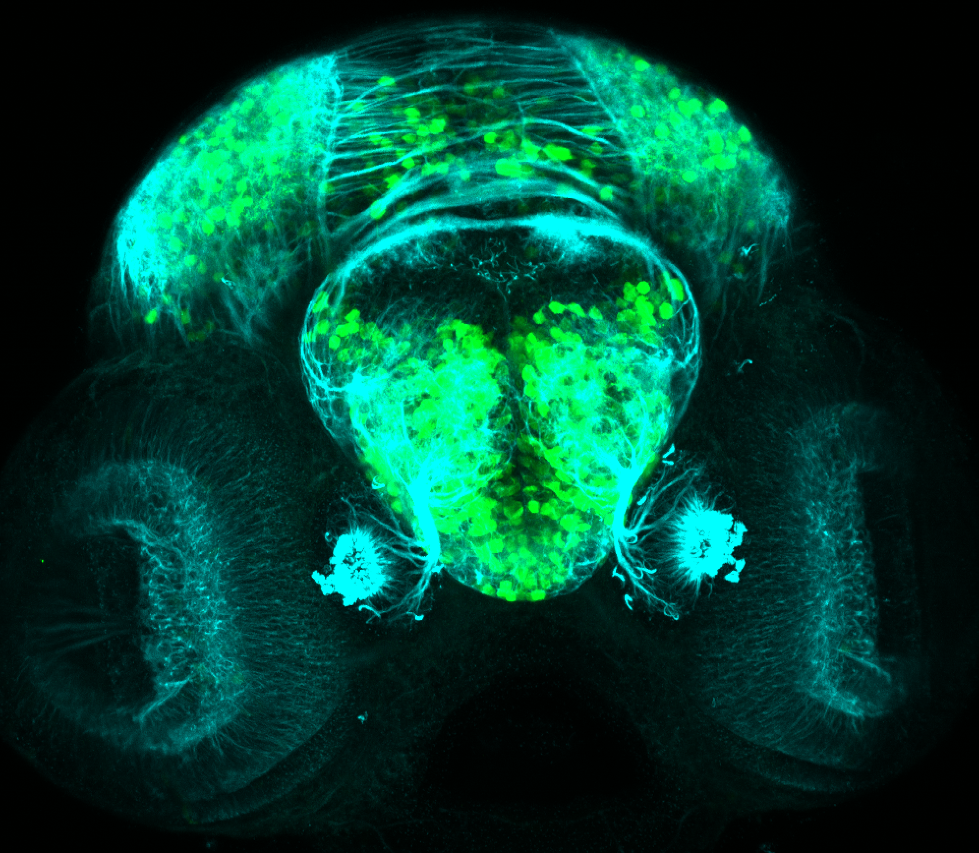
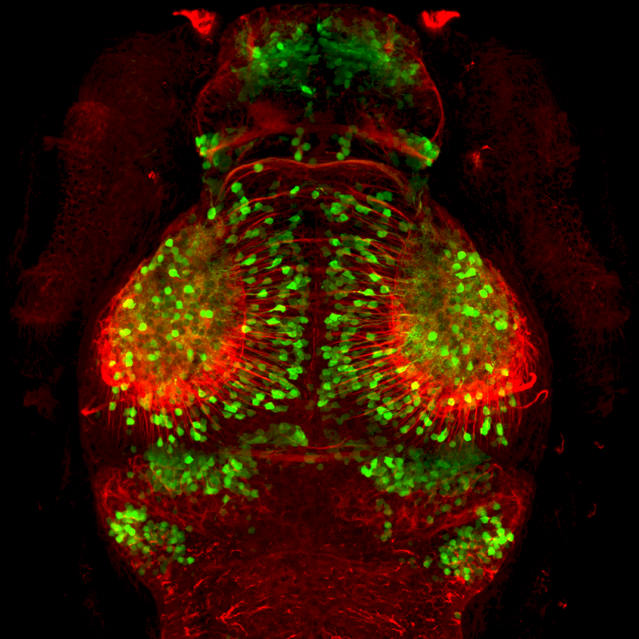
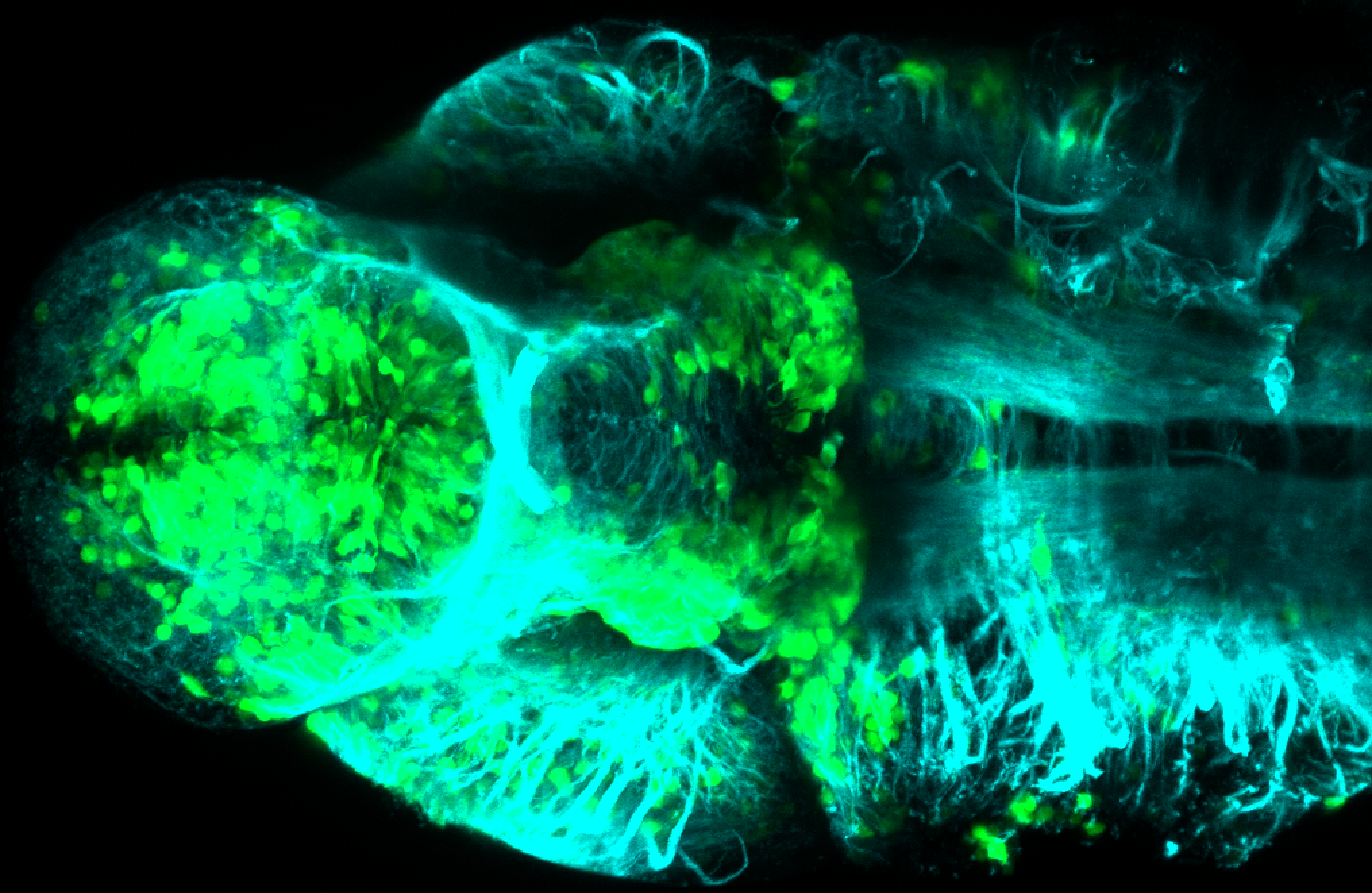
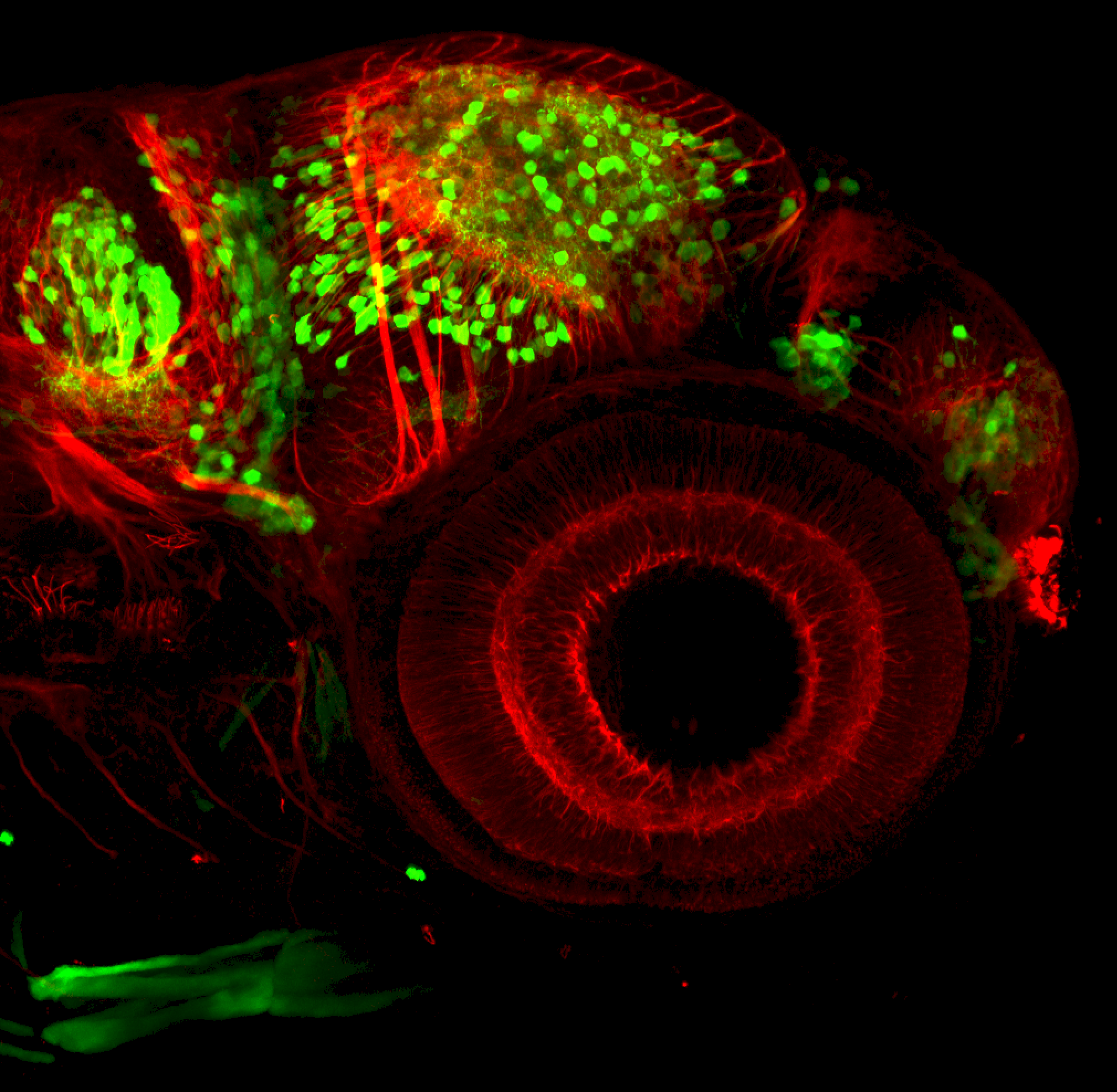
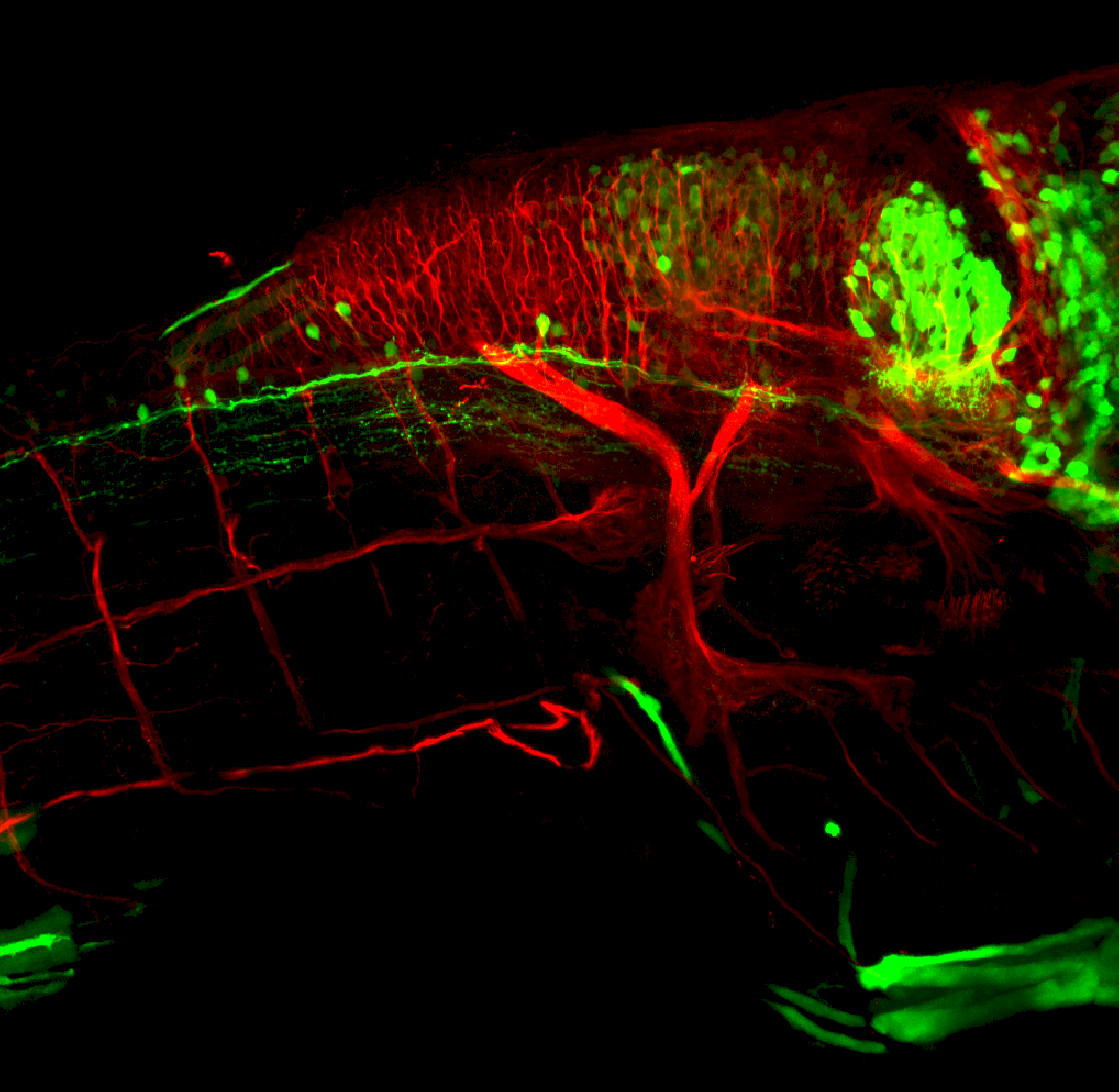
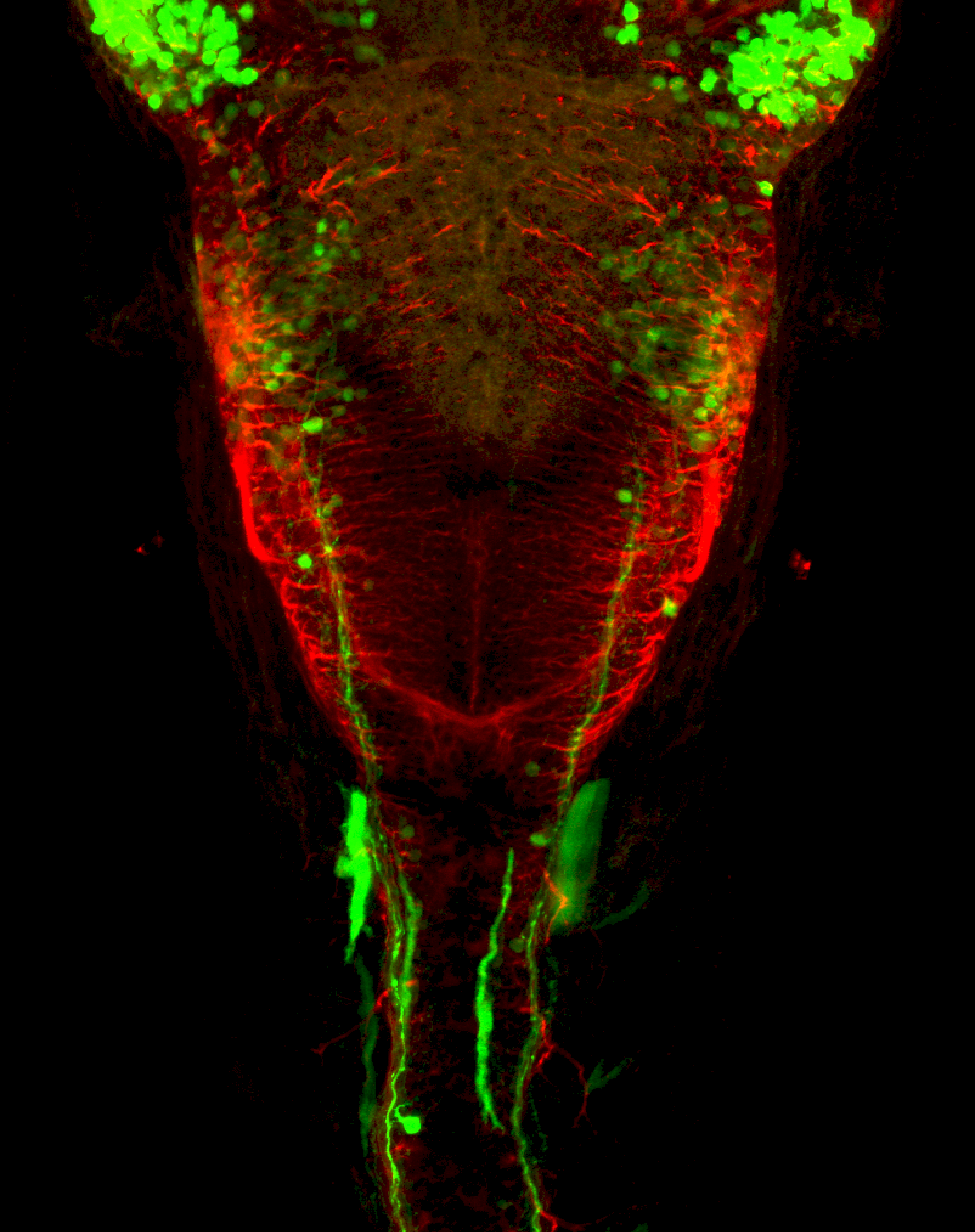
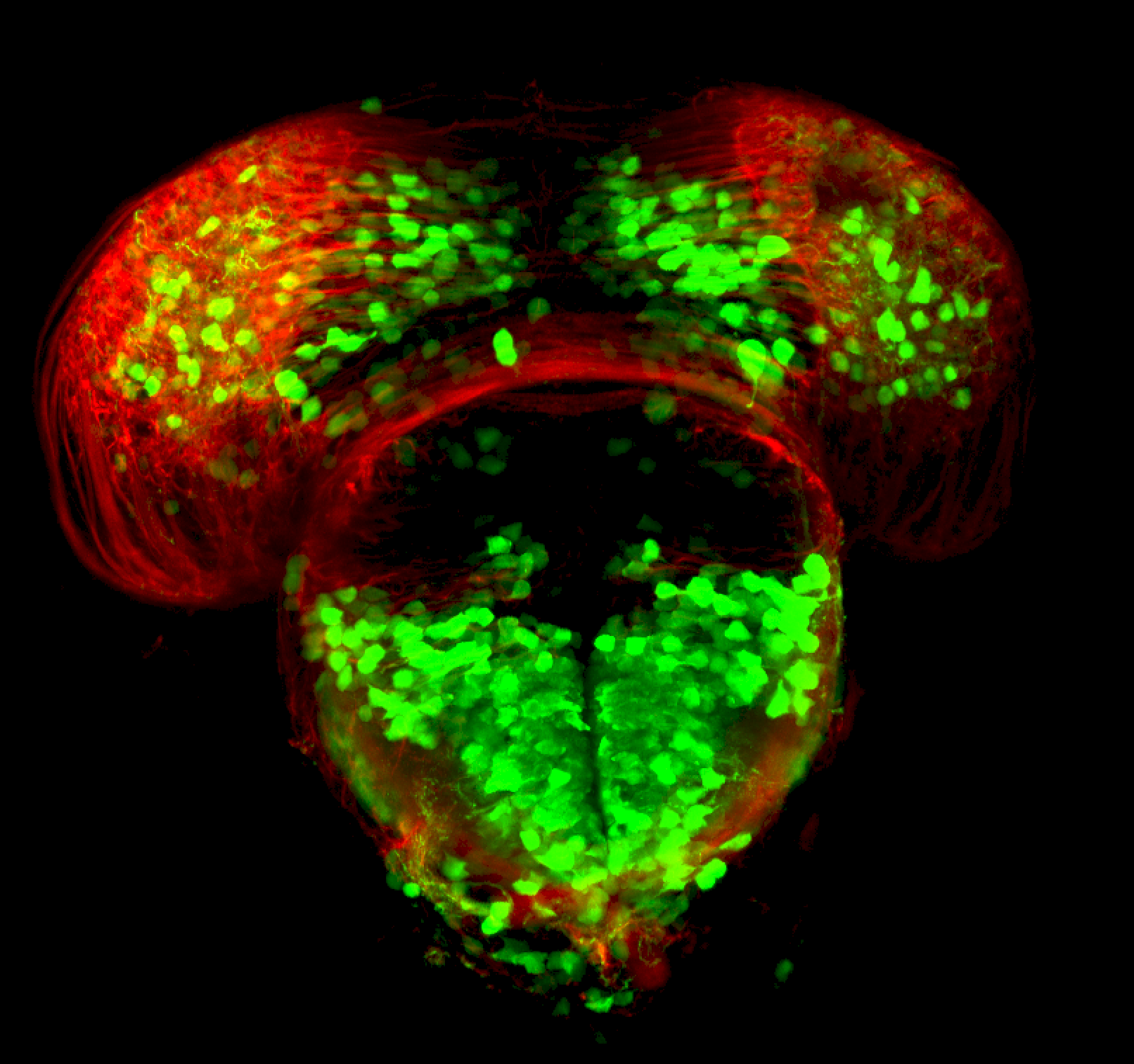
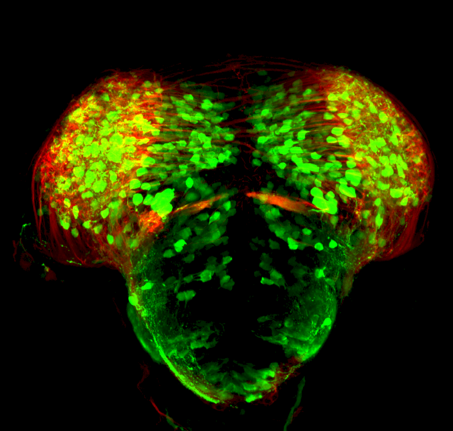
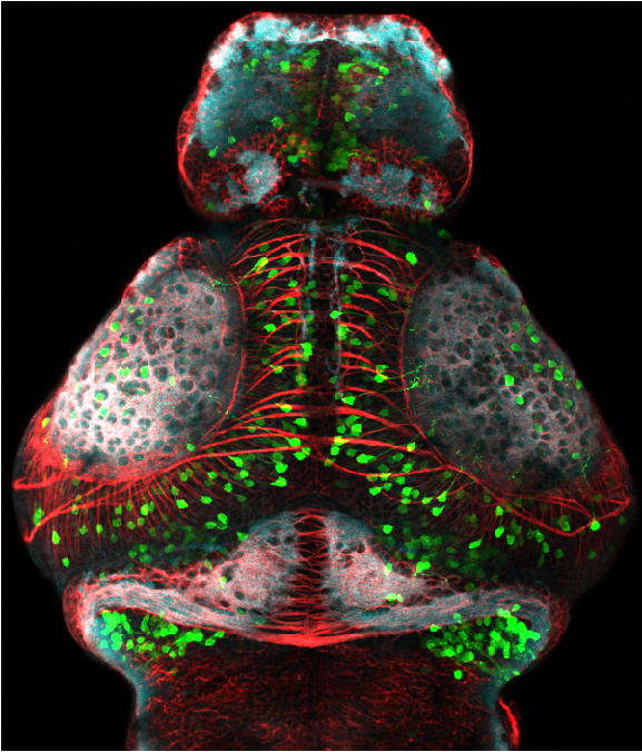
Tg(1.4dlx5a-dlx6a:GFP)ot1 larvae express GFP in subpallial neurons γ-aminobutyric acid (GABA)-expressing neurons. Several other brain regions also show GFP expression in this transgenic line such as the optic tectum and cerebellum.
Dlx homeobox genes play essential roles in the differentiation, migration and survival of subpallial precursor cells that will later give rise to diverse subtypes of γ-aminobutyric acid (GABA)-expressing neurons. They also participate in the regulation of the Gad genes encoding the enzymes necessary for GABA synthesis (Yu et al., 2011).
Mouse over the different areas of GFP expression in the interactive images below to see the name of the brain area.
More images
![Tg(1.4dlx5a-dlx6a:GFP)ot1 labelled with GFP, tubulin and sv2](https://images.squarespace-cdn.com/content/v1/58065fb61b631b37ff3ce66a/1531407473998-WQUH78JTGES40UOXEZ3A/Dlx+master.jpg)
![Lateral view of a 4dpf Tg(1.4dlx5a-dlx6a:GFP)ot1](https://images.squarespace-cdn.com/content/v1/58065fb61b631b37ff3ce66a/1531409819249-DO8CCF05176S3KP3KBQ5/dlx+tub+4dpf+lat.png)
![Lateral view of a 4dpf Tg(1.4dlx5a-dlx6a:GFP)ot1](https://images.squarespace-cdn.com/content/v1/58065fb61b631b37ff3ce66a/1531409817431-5N0JUPHW0S0MIW7L7OXG/dlx+4dpf+lat.png)
![Ventral view of a 4dpf Tg(1.4dlx5a-dlx6a:GFP)ot1](https://images.squarespace-cdn.com/content/v1/58065fb61b631b37ff3ce66a/1531410130181-GO1DXGPODTL3BN4UFWGX/dlx+4dpf+vent.png)
![Ventral view of a 4dpf Tg(1.4dlx5a-dlx6a:GFP)ot1 larvae](https://images.squarespace-cdn.com/content/v1/58065fb61b631b37ff3ce66a/1531410132006-HAVO62SEKBQ2HY2ZWJ1C/dlx+tub+4dpf+vent.png)
![Dorsal view of a 4dpf Tg(1.4dlx5a-dlx6a:GFP)ot1 larvae](https://images.squarespace-cdn.com/content/v1/58065fb61b631b37ff3ce66a/1531410248850-H9K8LRUKASPQLLOJ6A6H/dlx+4dpf+tub+dorsal.png)
![Dorsal view of a 4dpf Tg(1.4dlx5a-dlx6a:GFP)ot1 larvae](https://images.squarespace-cdn.com/content/v1/58065fb61b631b37ff3ce66a/1531410246671-FJPJSR8MOWPUATDZLSJR/dlx+4dpf+dorsal.png)
![Olfactory glomeruli in Tg(1.4dlx5a-dlx6a:GFP)ot1 labelled with GFP, tubulin and sv2](https://images.squarespace-cdn.com/content/v1/58065fb61b631b37ff3ce66a/1547652353042-EWI44HN3PGMRWPCIX4QL/4dpf+frontal+GMC.png)
![24hpf dorsal view of dlx:GFP with tubulin](https://images.squarespace-cdn.com/content/v1/58065fb61b631b37ff3ce66a/1547652443303-THORVLWEB40VWUB9HFGR/dlx+24hpf.png)
![Olfactory bulb Tg(1.4dlx5a-dlx6a:GFP)ot1 labelled with GFP, tubulin and sv2](https://images.squarespace-cdn.com/content/v1/58065fb61b631b37ff3ce66a/1547652349181-7R9DG64DPGTCGIRZ6DBF/dlx+4dpf+frontal+OB2.png)
![3dpf frontal view of dlx:GFP larvae](https://images.squarespace-cdn.com/content/v1/58065fb61b631b37ff3ce66a/1547652443185-52EIUGNKWO38QAS0F3IR/dlx+3dpf+frontal+GC.png)
![3dpf dorsal view dlx:GFP](https://images.squarespace-cdn.com/content/v1/58065fb61b631b37ff3ce66a/1547652450317-B1GE349MCP7RK780PNA3/dlx+3dpf+dorsal.png)
![2dpf dlx:GFP ventral view](https://images.squarespace-cdn.com/content/v1/58065fb61b631b37ff3ce66a/1547652454705-OMQJC8QKP0MC7C55UUVY/dlx+48hpf+vent.png)
![3dpf lateral view of dlx:GFP in forebrain](https://images.squarespace-cdn.com/content/v1/58065fb61b631b37ff3ce66a/1547652461548-A0Y6CIWSI9WF40FRQ8T4/dlx+3dpf+lat+FB+.png)
![3dpf late view of dlx:GFP hindbrain](https://images.squarespace-cdn.com/content/v1/58065fb61b631b37ff3ce66a/1547652466708-4T9ZXLND15SX0G6VOPD8/dlx+3dpf+HB+lat.png)
![3dpf dorsal view of dlx:GFP hindbrain](https://images.squarespace-cdn.com/content/v1/58065fb61b631b37ff3ce66a/1547652467741-9NO3QDCASON1UMBUG7B2/dlx+3dpf+hb.png)
![3dpf frontal section of dlx:GFP subpallium](https://images.squarespace-cdn.com/content/v1/58065fb61b631b37ff3ce66a/1547652472197-IEJHKIIL810OAZ6JVSIO/dlx+48hpf+frontal+section+2.png)
![3dpf thick section of dlx:GFP through midbrain/diencephalon](https://images.squarespace-cdn.com/content/v1/58065fb61b631b37ff3ce66a/1547652476889-V0GR5R44YRJ7EJXFVOB0/dlx+48hpf+frontal+section.png)
Expressed in:
olfactory bulb, subpallium, pallium, preoptic area, prethalamus, posterior tuberculum, hypothalamus, optic tectum, cerebellum.
Key Publications
Zerucha, T., Stuhmer, T., Hatch, G., Park, B.K., Long, Q., Yu, G., Gambarotta, A., Schultz, J.R., Rubenstein, J.L., and Ekker, M. (2000)
A highly conserved enhancer in the Dlx5/Dlx6 intergenic region is the site of cross-regulatory interactions between dlx genes in the embryonic forebrain.
The Journal of neuroscience : the official journal of the Society for Neuroscience. 20(2):709-721.
Yu, M., Xi, Y., Pollack, J., Debiais-Thibaud, M., Macdonald, R.B., and Ekker, M. (2011)
Activity of dlx5a/dlx6a regulatory elements during zebrafish GABAergic neuron development.
Int. J. Dev. Neurosci.. 29(7):681-91.