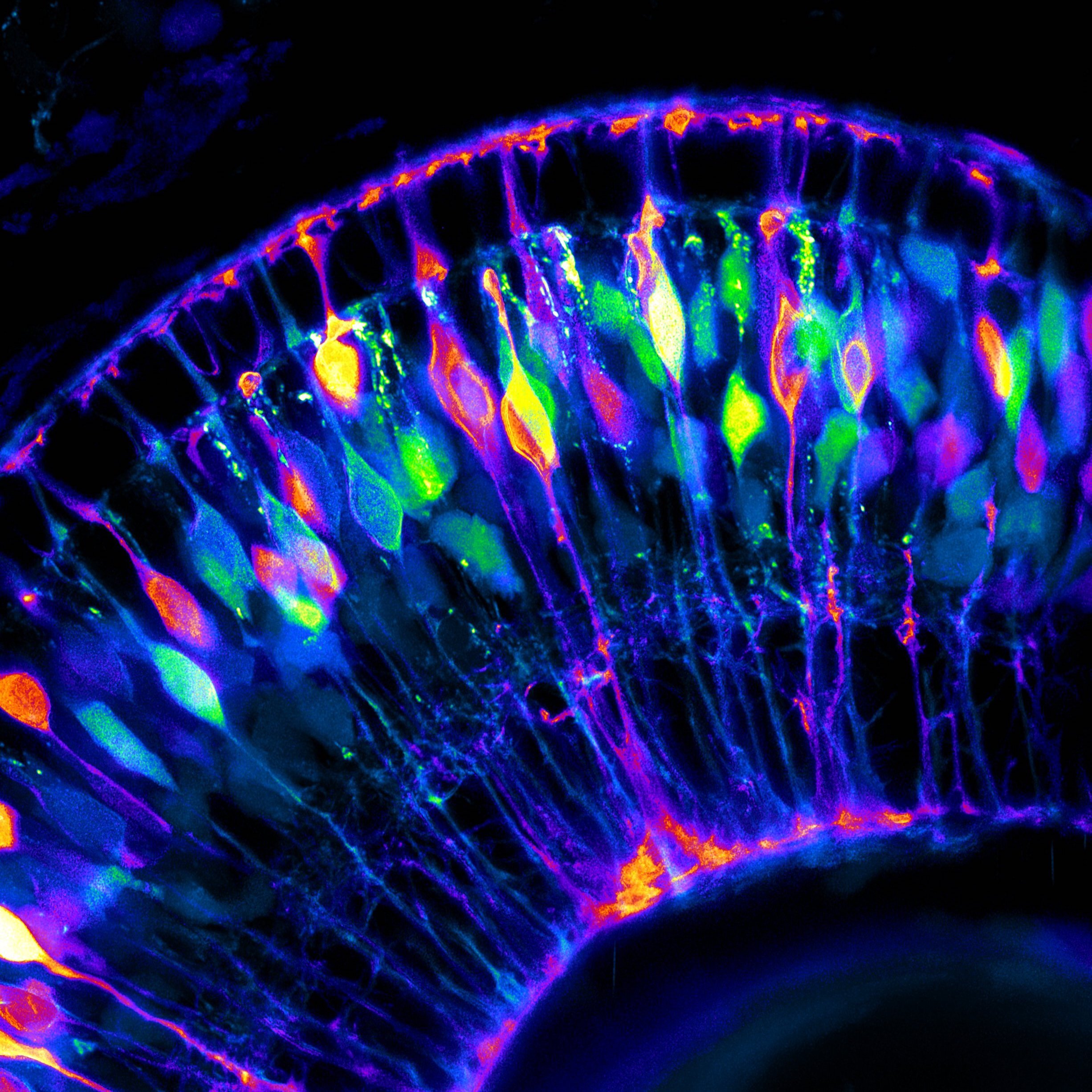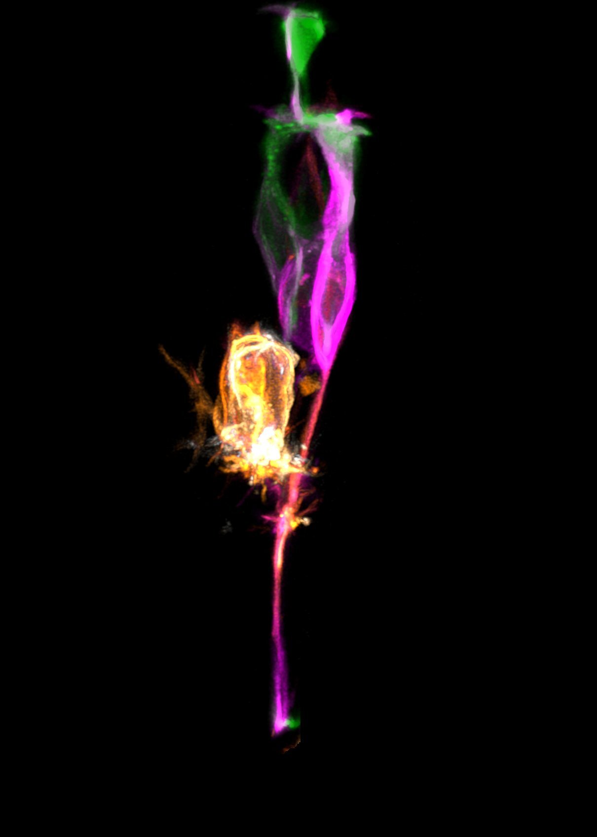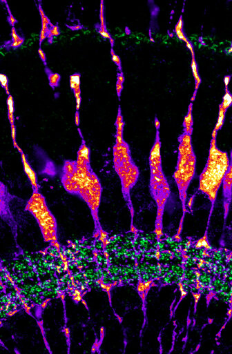
![Image by Xhuljana Durmishi](https://images.squarespace-cdn.com/content/v1/58065fb61b631b37ff3ce66a/1658414247554-3D5DOU89X4923F1LC68Q/1-72hpf_TP1eGFP-CAAX.jpg)
Image by Xhuljana Durmishi
![Section of 96 hpf zerbafish retina Tg(tp1:Venus) anti-Ca1 in green, anti-zrf1 in red and DAPI in blue](https://images.squarespace-cdn.com/content/v1/58065fb61b631b37ff3ce66a/1695991078215-F4H9GQWI0TD3HUS3YOQ0/96hpf+F1+Carbonic+anhydrase+488+zrf1-550+Tp1Venus-1.png)
Section of 96 hpf zerbafish retina Tg(tp1:Venus) anti-Ca1 in green, anti-zrf1 in red and DAPI in blue
Image by Aanandita Korthurkar
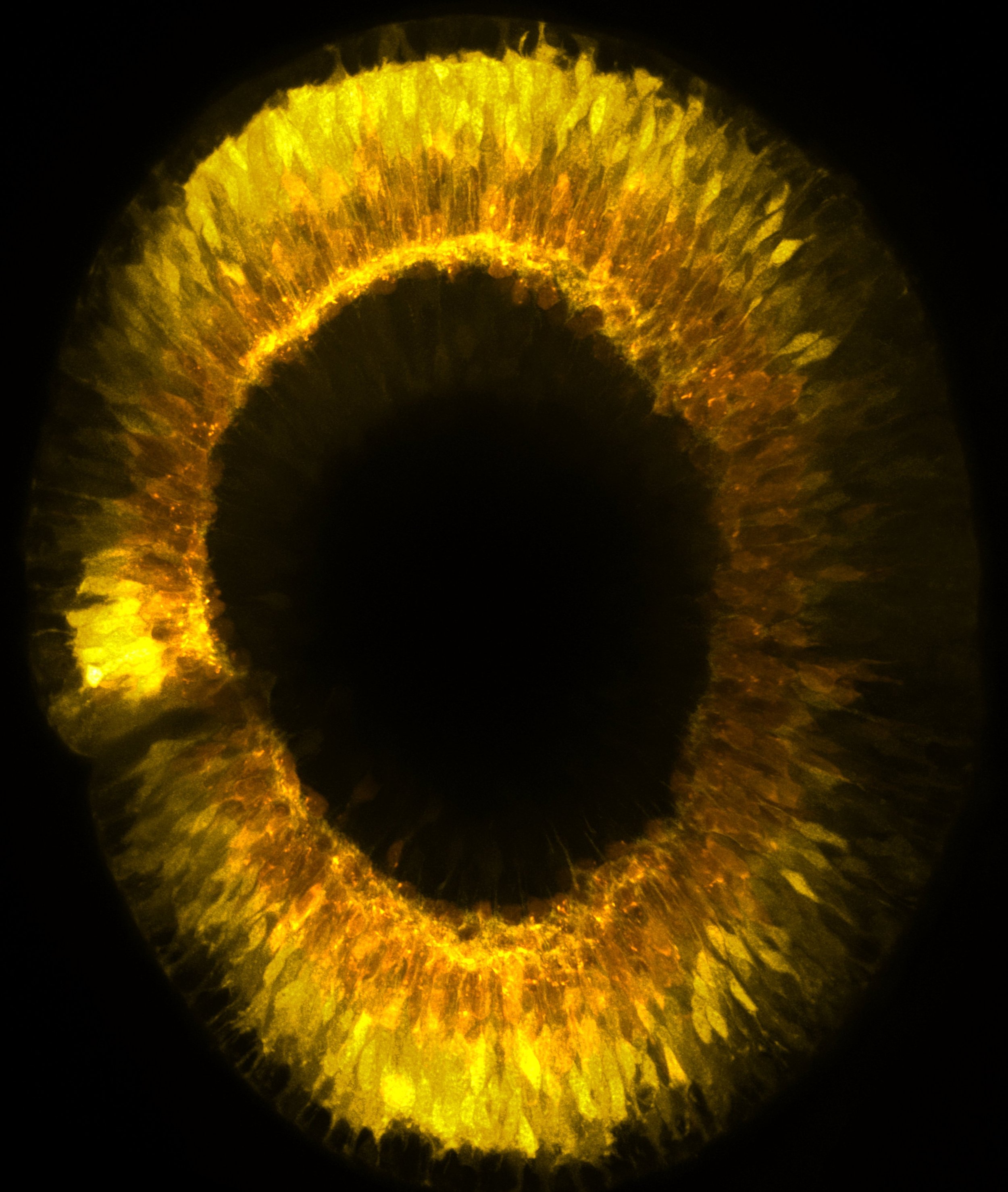
![By Xhuljana Durmishi](https://images.squarespace-cdn.com/content/v1/58065fb61b631b37ff3ce66a/1658414266419-BD37Q5ZEF86KFUGTN59T/4-48hpf_vsx1-ptf1a.jpg)
By Xhuljana Durmishi
![Image by Gregory Patient A three round IBEX on a five days post fertilisation zebrafish retinal section, showing nuclei, different photoreceptor components, Müller glia, microglia, amacrine and retinal ganglion cells. The markers are: green – Gluta](https://images.squarespace-cdn.com/content/v1/58065fb61b631b37ff3ce66a/1678463460744-ENHF9T27QZTHAIXBI6LE/image-asset.png)
Image by Gregory Patient
A three round IBEX on a five days post fertilisation zebrafish retinal section, showing nuclei, different photoreceptor components, Müller glia, microglia, amacrine and retinal ganglion cells. The markers are: green – Glutamate Synthetase, red – GNAT2, cyan – M/L opsin, white – HuC/D, yellow – PNA lectin, magenta – LCP1, blue – DAPI.
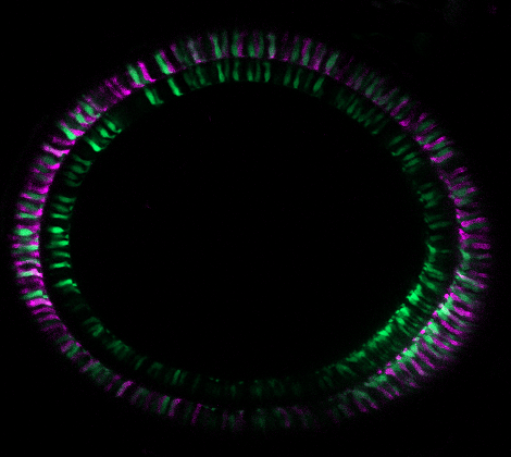
![Image by Manuela Lahne](https://images.squarespace-cdn.com/content/v1/58065fb61b631b37ff3ce66a/1674596371034-L761XDRK4UYTE6ZL1LOY/Manuela+3.png)
Image by Manuela Lahne
![Section cell body.jpg](https://images.squarespace-cdn.com/content/v1/58065fb61b631b37ff3ce66a/1695721946747-6K1IGZPWDZPZN974LIQV/Section+cell+body.jpg)
Section cell body.jpg
Image by Nicole Noel
Section of cell body of killifish retina
![Glutamine synthetase](https://images.squarespace-cdn.com/content/v1/58065fb61b631b37ff3ce66a/1561391286298-9Y40FUHBW8YWJ54NSJI2/Publications.jpg)
Glutamine synthetase
Image by Elisabeth Kugler
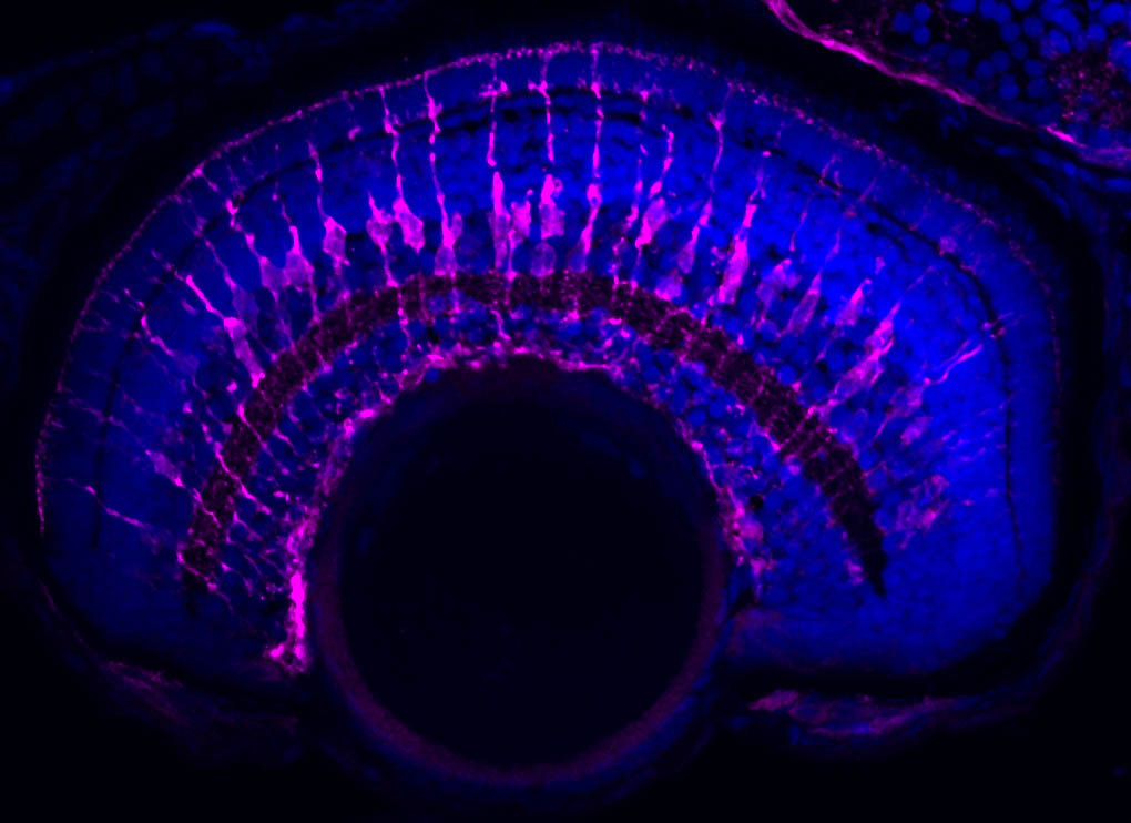
Müller glia express specific proteins that allow them to carry out their support functions in the retina. Cryosection of the zebrafish retina and immunohistochemistry for glutamine synthetase (magenta).
![Image by Ryan MacDonald](https://images.squarespace-cdn.com/content/v1/58065fb61b631b37ff3ce66a/1671201881984-FYV17ZE0340O6341ZL4X/MullerGlia-AC.png)
Image by Ryan MacDonald
![By Xhuljana Durmishi](https://images.squarespace-cdn.com/content/v1/58065fb61b631b37ff3ce66a/1658414275310-S0D3Y7K30OY9SQ5ZZB6L/6-48hpf_vsx1-ptf1a.jpg)
By Xhuljana Durmishi
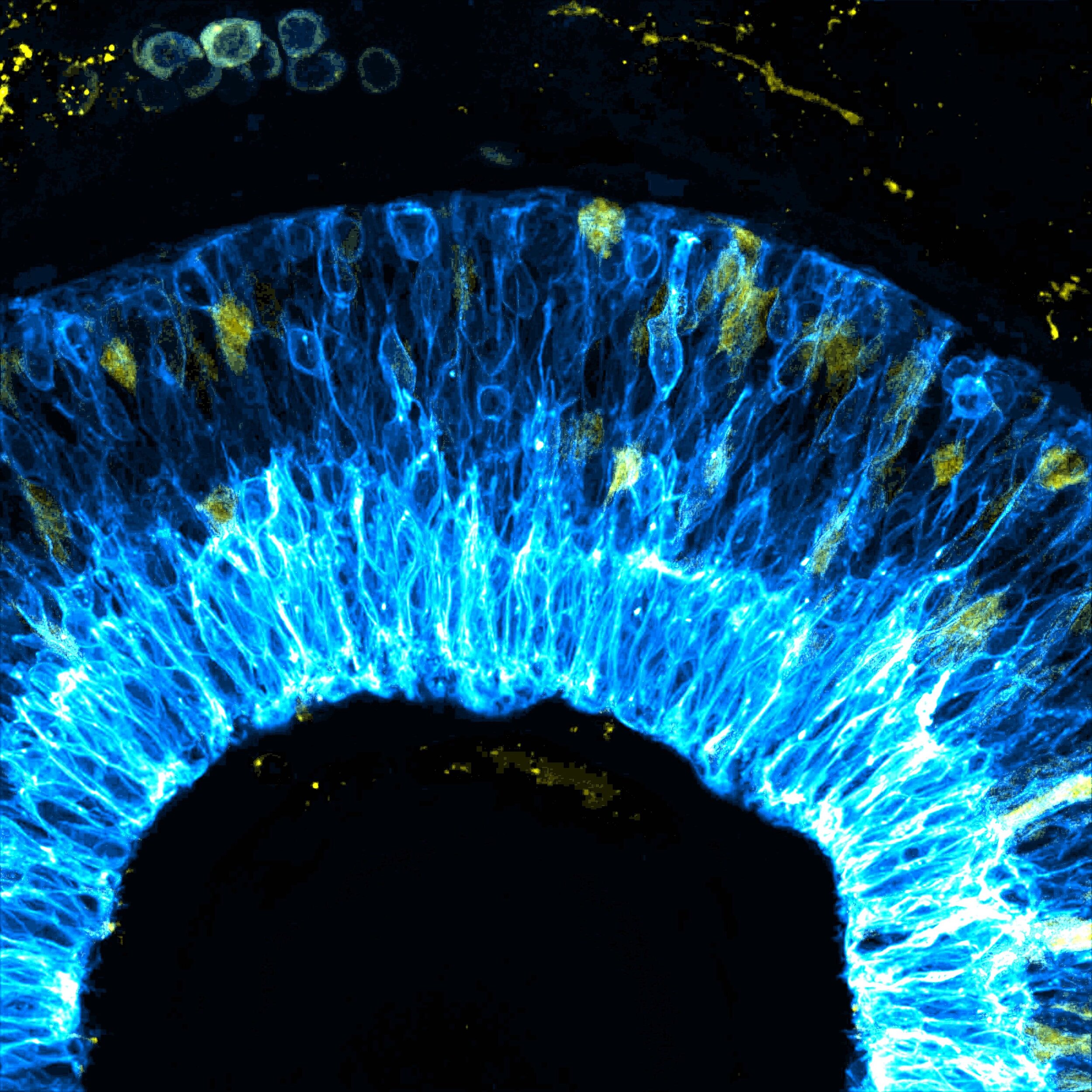
![Image by Elisabeth Kugler False-colour illustration of zebrafish retina neurons (blue) and glia progenitors (yellow) at 48 hours post fertilization, acquired with Zeiss AiryScan microscopy. Image processing was conducted with Fiji software.](https://images.squarespace-cdn.com/content/v1/58065fb61b631b37ff3ce66a/1612265628387-HIHJYVKPCZCMGNJKGWAH/Composite.jpg)
Image by Elisabeth Kugler
False-colour illustration of zebrafish retina neurons (blue) and glia progenitors (yellow) at 48 hours post fertilization, acquired with Zeiss AiryScan microscopy. Image processing was conducted with Fiji software.
![Tg(gfap:gfp)](https://images.squarespace-cdn.com/content/v1/58065fb61b631b37ff3ce66a/1561557824533-DFUWXIV5D01XPDFUX14M/Picture+2+%281%29.jpg)
Tg(gfap:gfp)
Image by Elisabeth Kugler
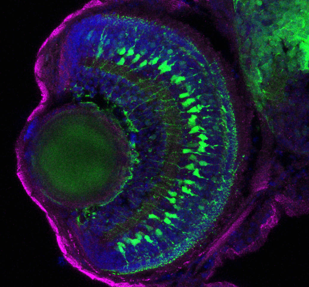
Müller glia are specifically labelled in the transgenic background Tg(gfap:gfp). We can use transgenic zebrafish or antibodies to specifically label and identify the glia or neurons in the retina. Cryosection of the zebrafish retina and immunohistochemistry for GFP.
![Müller glia and retinal neurons](https://images.squarespace-cdn.com/content/v1/58065fb61b631b37ff3ce66a/1657562192266-IN3CCEW12ND3X4O0N2PY/website+pic+2.png)
Müller glia and retinal neurons
Image by Natalia Jaroszynska
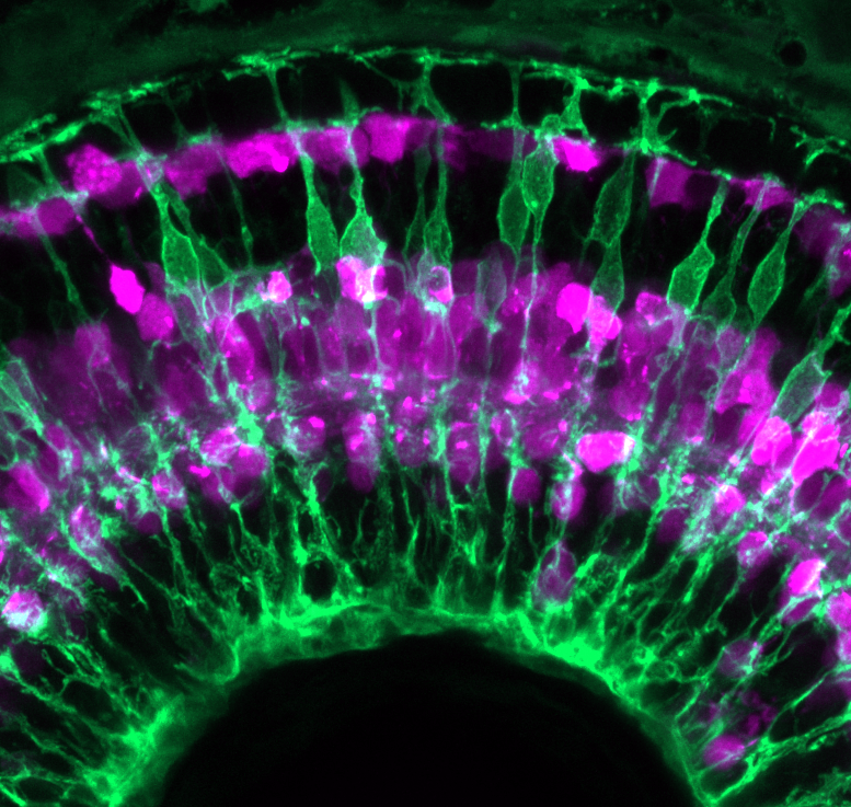
Müller glia (green) interact with all of the surrounding retinal cells in different retinal layers, including horizontal and amacrine cells (magenta). Zebrafish retina at 3 days post fertilisation.
![Glial cells support healthy neurons](https://images.squarespace-cdn.com/content/v1/58065fb61b631b37ff3ce66a/1561391211363-TXPDAGTWDOGUEALGIOD9/Alternate+2.jpg)
Glial cells support healthy neurons
By Elisabeth Kugler
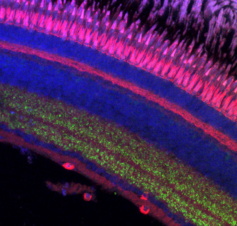
As the zebrafish retina matures glial cells must support the tissue to maintain healthy neurons and their connections or synapses. Cryosection of the adult zebrafish retina and staining with DAPI (blue), phalloidin (red), zpr3 (photoreceptors; magenta) and ribeye (synapses; green).
![The retina connects to the visual centres in the brain](https://images.squarespace-cdn.com/content/v1/58065fb61b631b37ff3ce66a/1561391215257-VZSYUQAP98ZUCO2IE2LV/Alternate1.jpg)
The retina connects to the visual centres in the brain
Image by Elisabeth Kugler
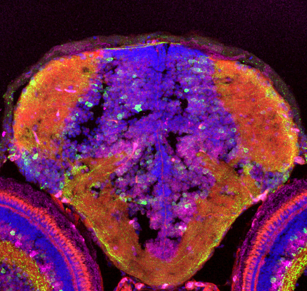
The retina connects to the visual centres in the brain. Cryosection of the zebrafish brain and staining with DAPI (blue), phalloidin (red), HuC/D (neurons; magenta) and GFP (green). Genotype is Tg(NBT-GCaMP3) which labels neurons in the retina and tectum.
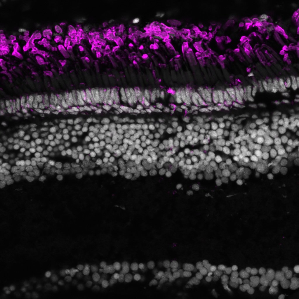
![202305 zpr-1-magenta_DAPI-cyan_12wo-killifish.jpg](https://images.squarespace-cdn.com/content/v1/58065fb61b631b37ff3ce66a/1695721684502-UTSOCJYZSI1GTN0B0RYY/202305+zpr-1-magenta_DAPI-cyan_12wo-killifish.jpg)
202305 zpr-1-magenta_DAPI-cyan_12wo-killifish.jpg
Image by Nicole Noel
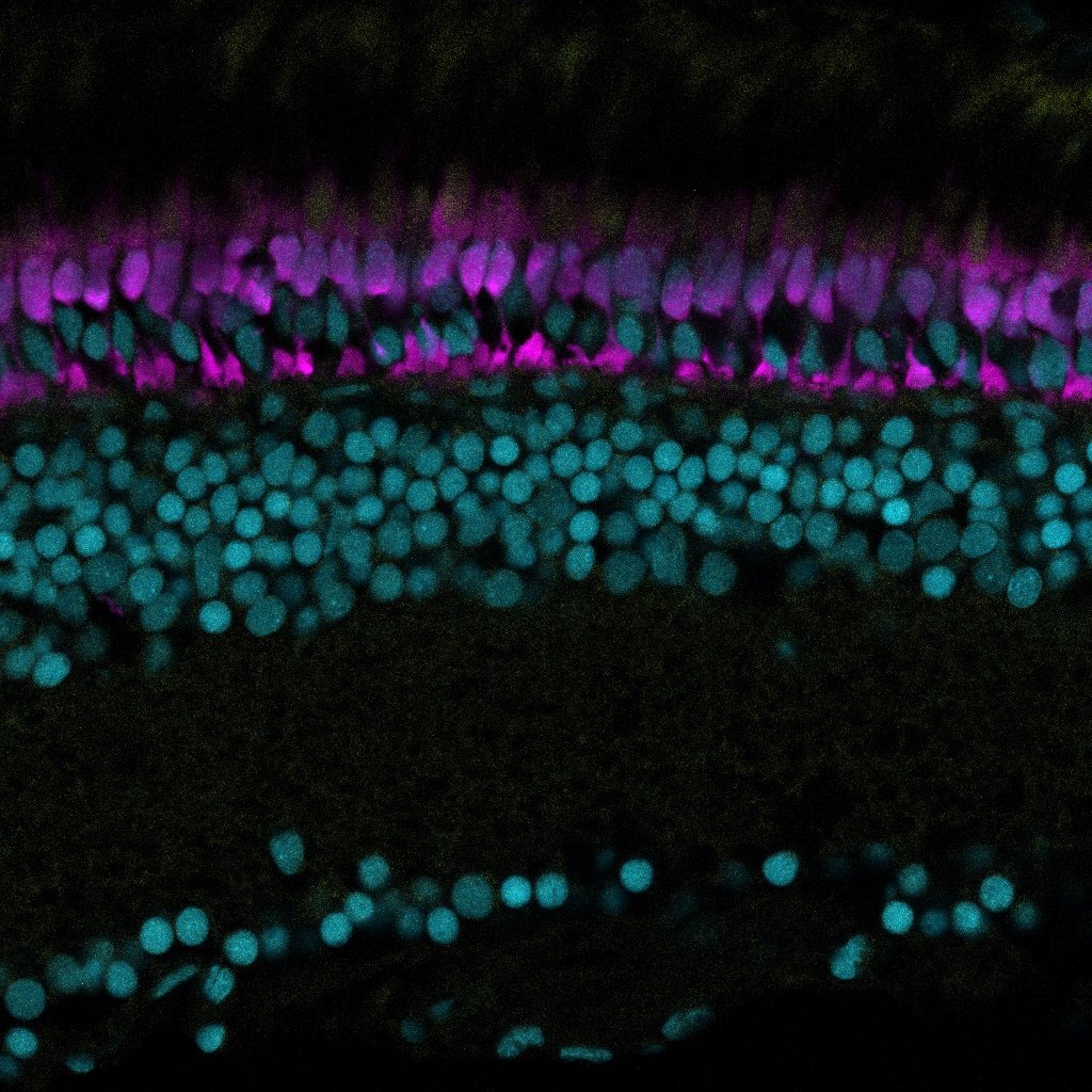
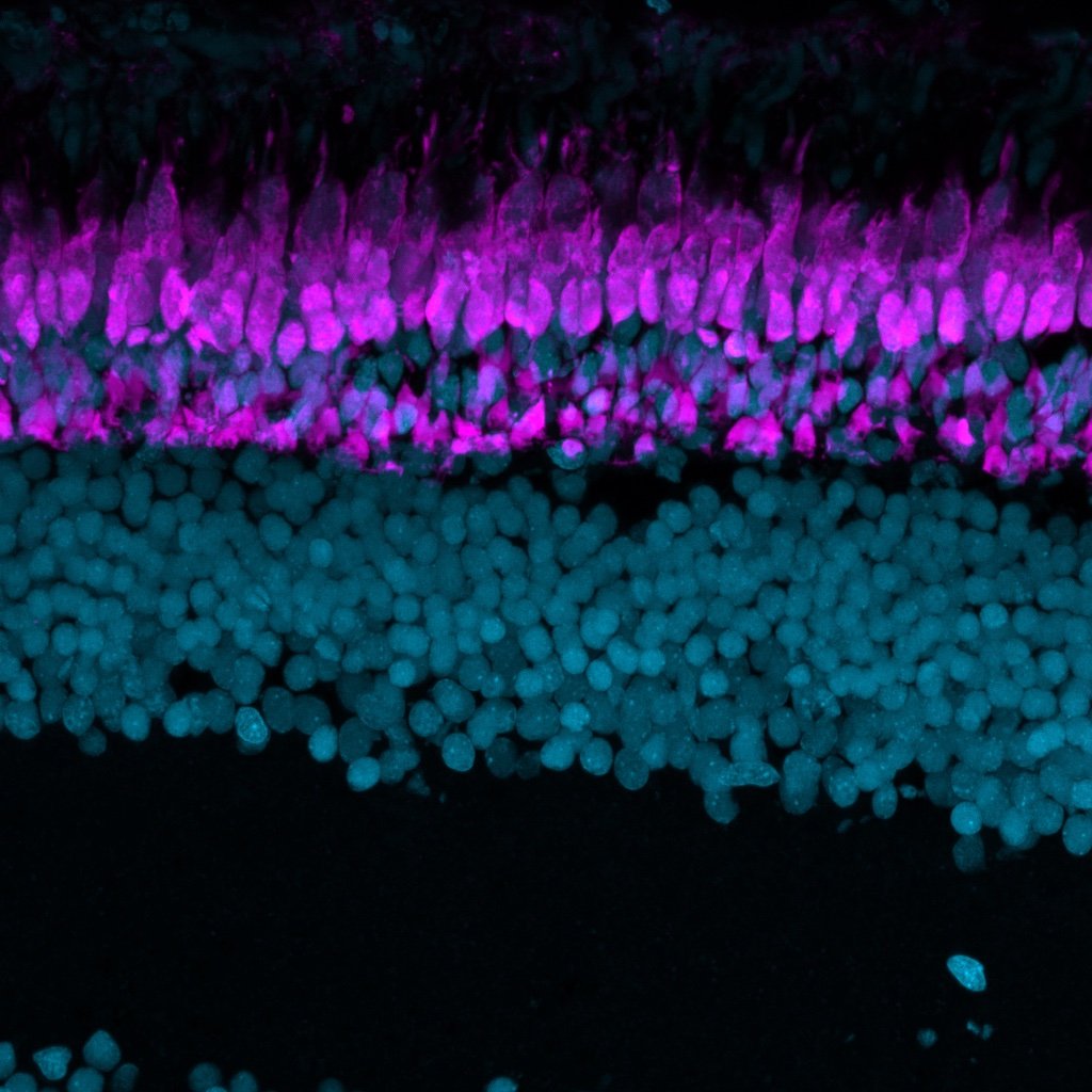
Immunohistochemistry on 12 wpf killifish retina section. Magenta is zpr-1 and DAPI is in cyan.
![Semi-super resolution reconstruction of Müller glia cells](https://images.squarespace-cdn.com/content/v1/58065fb61b631b37ff3ce66a/1561557298181-FJAGIYVZH1RLU8GZQ3SO/Alternate5.jpg)
Semi-super resolution reconstruction of Müller glia cells
Image by Elisabeth Kugler
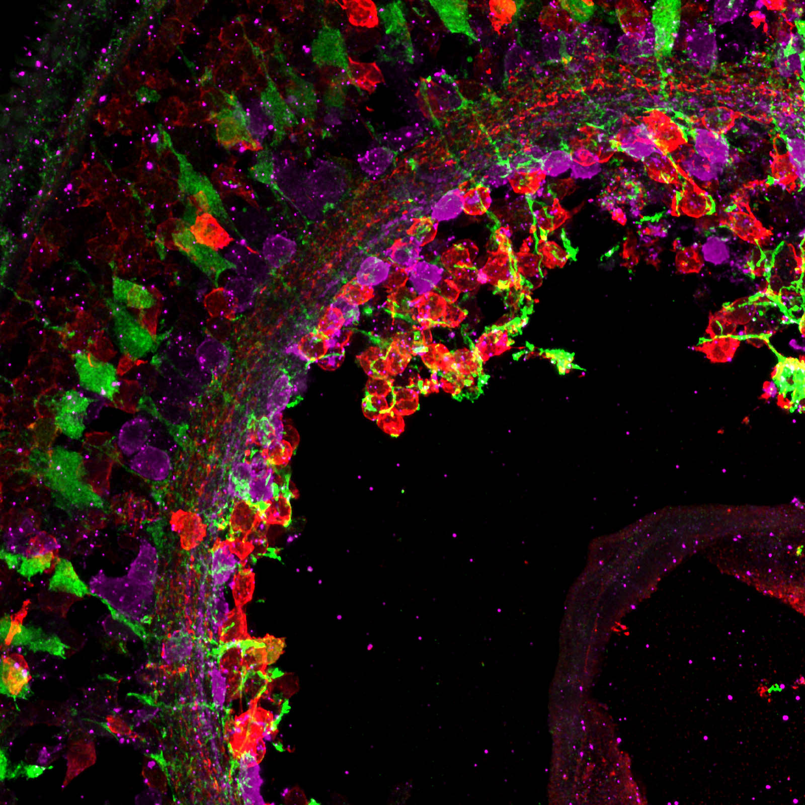
We are trying to push the limits of our imaging to understand glial cells. This is a semi-super resolution reconstruction of Müller glia cells (green) and two different classes of amacrine cells (magenta and red) that will come together to make distinct synapses in the inner plexiform layer. Cryosection of the zebrafish retina taken on a Leica Sp8 and processed with Lightning software.
![HCR in a 4-day old zebrafish transgenic (tp1:Venus) retina, showing Vsx1 (magenta) mRNA](https://images.squarespace-cdn.com/content/v1/58065fb61b631b37ff3ce66a/1657561390038-B5R7WW6HBB1EAPTL4IXF/website+pic.png)
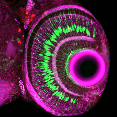
HCR in a 4-day old zebrafish transgenic (tp1:Venus) retina, showing Vsx1 (magenta) mRNA
Image by Natalia Jaroszynska
![Muller glia in the zebrafish retina](https://images.squarespace-cdn.com/content/v1/58065fb61b631b37ff3ce66a/1612265619481-8JIBTDTG0ZJCA1NT6VN6/001.png)
Muller glia in the zebrafish retina
Image by Ryan MacDonald
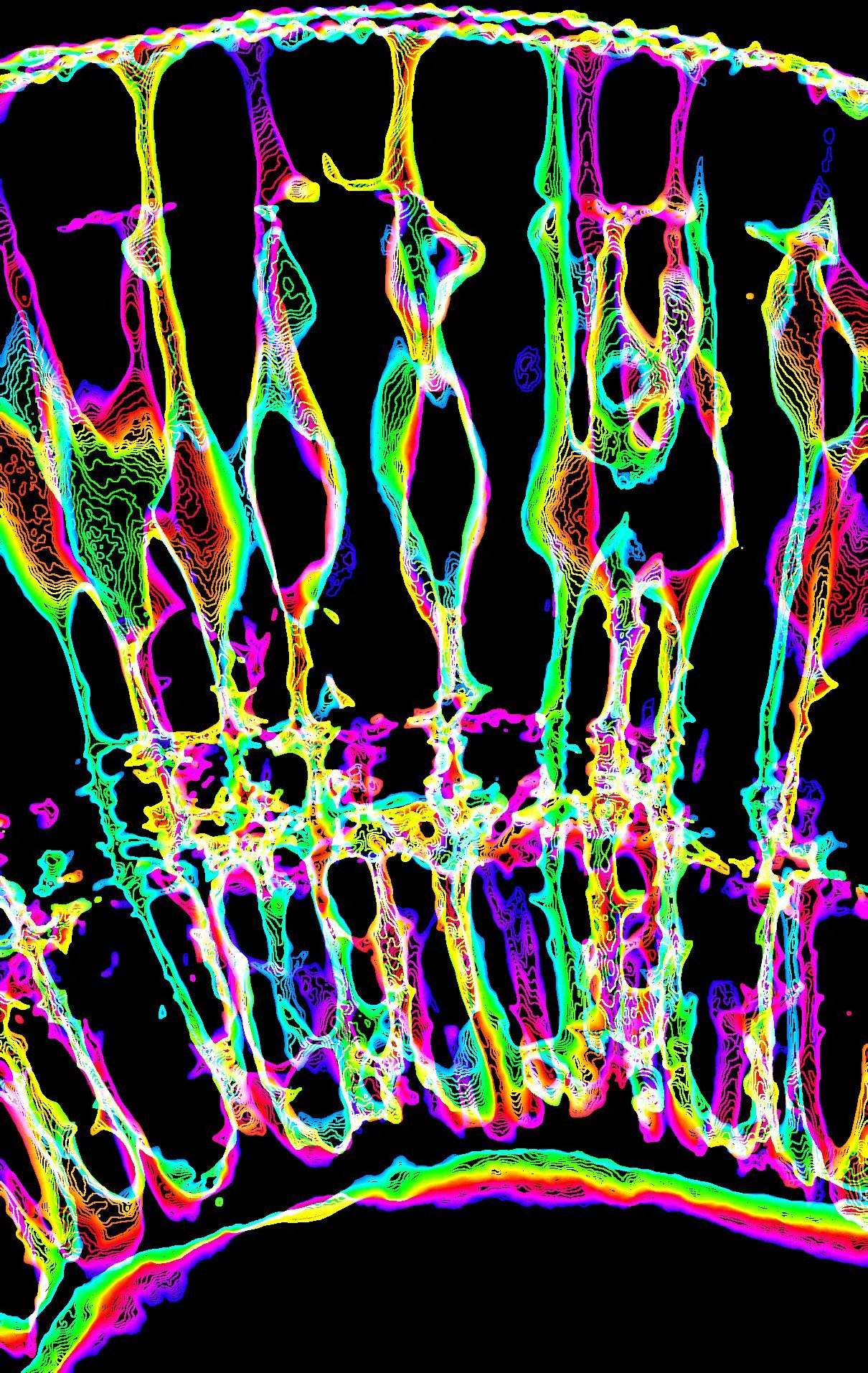
False-colour illustration of zebrafish retina Muller glia cells at 4dpf; same image reproduced with different look up tables. Image processing was conducted with Fiji software.
![Image by Xhuljana Durmishi](https://images.squarespace-cdn.com/content/v1/58065fb61b631b37ff3ce66a/1658414252874-JBCUENJFAIPNBKW5IRFF/2-72hpf_vsx1-ptf1a.jpg)
Image by Xhuljana Durmishi
![zpr-3 killifish.jpg](https://images.squarespace-cdn.com/content/v1/58065fb61b631b37ff3ce66a/1695728855420-70L5SDFA6EX2VBBAFE1T/zpr-3+killifish.jpg)
zpr-3 killifish.jpg
Image by Nicole Noel
Section of killifish retina zpr-3 in magenta
![Image by Elisabeth Kugler Image shows multi-colour false segmentation of the zebrafish retina at 3dpf. Image processing was conducted with Fiji software.](https://images.squarespace-cdn.com/content/v1/58065fb61b631b37ff3ce66a/1612265630739-F36DBAN7I0ZYMAUFY2V2/interactive+watershed-Z.jpg)
Image by Elisabeth Kugler
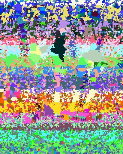
Image shows multi-colour false segmentation of the zebrafish retina at 3dpf. Image processing was conducted with Fiji software.
![Muller glia connections.](https://images.squarespace-cdn.com/content/v1/58065fb61b631b37ff3ce66a/1612265635300-Y32MCCZL3FNKJYU2ZW7J/MAX_z-reduced_60hpf_ath5gapGFP_cslmCherry_e1-01-Airyscan+Processing-482.jpg)
Muller glia connections.
Image by Elisabeth Kugler
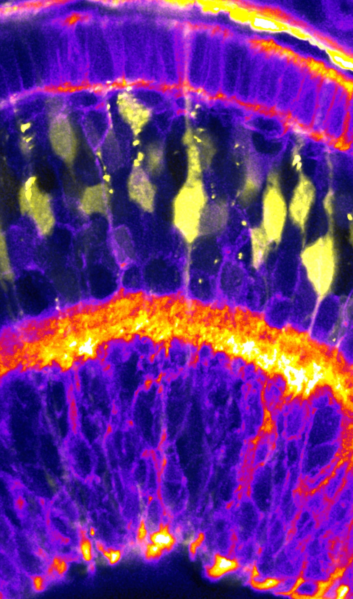
False-colour illustration of zebrafish retina neurons (blue) and glia progenitors (yellow) at 48 hours post fertilization, acquired with Zeiss AiryScan microscopy. Image processing was conducted with Fiji software.
![Between the eyes](https://images.squarespace-cdn.com/content/v1/58065fb61b631b37ff3ce66a/1657560940377-A9BHBUCWX06B35D8JEFH/Snap-542.jpg)
Between the eyes
Image by Natalia Jaroszynska
Immunostaining on 5 day old coronal zebrafish cryosections. Amacrine and retinal ganglion cells labelled with HuC/D (magenta) and bipolar cells labelled with PKC (red). Nuclei labelled with DAPI.
![Image by Gregory Patient A multiplex of four rabbit antibodies on the same five days post fertilisation zebrafish retinal section using micro-conjugation kits. Photoreceptors shown in green by GNAT2, Müller glia cells in magenta with RLBP1 and syna](https://images.squarespace-cdn.com/content/v1/58065fb61b631b37ff3ce66a/1678463653393-UHJPQ071IR2QZZ4M0LY1/image-asset.png)
Image by Gregory Patient
A multiplex of four rabbit antibodies on the same five days post fertilisation zebrafish retinal section using micro-conjugation kits. Photoreceptors shown in green by GNAT2, Müller glia cells in magenta with RLBP1 and synaptic terminals shown in yellow by PKCβ and in cyan with Ribeye.
![Muller glia support synapses](https://images.squarespace-cdn.com/content/v1/58065fb61b631b37ff3ce66a/1612265979583-HYXTOUS7J603A5I0GHQF/Screenshot+2020-10-30+at+11.32.15.png)
Muller glia support synapses
Image by Elisabeth Kugler
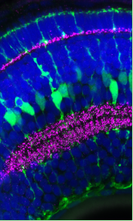
Zebrafish retinal synapses (magenta) and Muller glia (green) at 120 hours post fertilisation, acquired with Zeiss AiryScan microscopy. Image processing was conducted with Fiji software.
![Image by Manuela Lahne](https://images.squarespace-cdn.com/content/v1/58065fb61b631b37ff3ce66a/1674596363691-KGMAYAPGF71YZNWQVPOJ/manuela+2.png)
Image by Manuela Lahne
![Image by Elisabeth Kugler](https://images.squarespace-cdn.com/content/v1/58065fb61b631b37ff3ce66a/1629817340500-SQ08SU87E27KV9G5AHUL/FocalPlane_01.jpg)
Image by Elisabeth Kugler
![Image by Elisabeth Kugler](https://images.squarespace-cdn.com/content/v1/58065fb61b631b37ff3ce66a/1629817346649-0Z60N1A3XI7SSZ19BNM5/MAX_colored_60hpf.jpg)
Image by Elisabeth Kugler
![Image by Elisabeth Kugler](https://images.squarespace-cdn.com/content/v1/58065fb61b631b37ff3ce66a/1629817381551-6OTMM29WS5OKWP5WK1XG/Alicia.jpg)
Image by Elisabeth Kugler
![Image by Elisabeth Kugler](https://images.squarespace-cdn.com/content/v1/58065fb61b631b37ff3ce66a/1655046777737-NYHR75LB16PUTXCCOSCU/FU4-k2XX0AA4tU-.jpg)
Image by Elisabeth Kugler
![HCR in 5 dpf wildtype zebrafish retina, showing cyp26a1 mRNA](https://images.squarespace-cdn.com/content/v1/58065fb61b631b37ff3ce66a/1695991785527-S57QB3RQA4HCJQSTLGL0/MAX_17-5-23+Cdn+2+A-A+cyp26a1+B1+488+glula+b3+546-01.czi+-+17-5-23+Cdn+2+A-A+cyp26a1+B1+488+glula+b3+546-01.czi+%233-1.png)
HCR in 5 dpf wildtype zebrafish retina, showing cyp26a1 mRNA
Image by Aanandita Korthurkar
![Killifish retina](https://images.squarespace-cdn.com/content/v1/58065fb61b631b37ff3ce66a/1669983637847-3EV16ZWWSPRPXQM3P7P8/M_8+bits_Scale+bar_11.11.21+female+Killifish+retina+2+months+old+DAPI+GS+647+GFAP+488+40x+objective+speed+5s_Substack+%281-10%29.jpg)
Killifish retina
Image by Eva-Maria Breitenbach
It shows the the image of a 2 mpf female killifish with anti-Gs staining which labels Müller glia cells.
![Image by Isabel Bravo](https://images.squarespace-cdn.com/content/v1/58065fb61b631b37ff3ce66a/1671201882221-028LGFLLYFAZGE7CR312/MullerGlia.png)
Image by Isabel Bravo
![zpr-1 killifish 2.jpg](https://images.squarespace-cdn.com/content/v1/58065fb61b631b37ff3ce66a/1695728855123-VOUGISVNNA4JMD2KG0PI/zpr-1+killifish+2.jpg)
zpr-1 killifish 2.jpg
Image by Nicole Noel
Section of killifish retina zpr-1 in magenta and DAPI in cyan
![By Xhuljana Durmishi](https://images.squarespace-cdn.com/content/v1/58065fb61b631b37ff3ce66a/1658414255606-CXU3MQPZWBBZADGF2I9U/3-72hpf_TP1Venus_immuno.jpg)
By Xhuljana Durmishi
![By Xhuljana Durmishi](https://images.squarespace-cdn.com/content/v1/58065fb61b631b37ff3ce66a/1658414269755-XAE49TMDUDK2RX9GMMWH/5-72hpf_TP1-krdl.jpg)
By Xhuljana Durmishi
![Actin cytoskeleton of Müller glia](https://images.squarespace-cdn.com/content/v1/58065fb61b631b37ff3ce66a/1675691195090-G8V8WHP0IJD68J7D29HC/MG+actin+cytoskeleton.png)
Actin cytoskeleton of Müller glia
Image by Natalia Jaroszynska
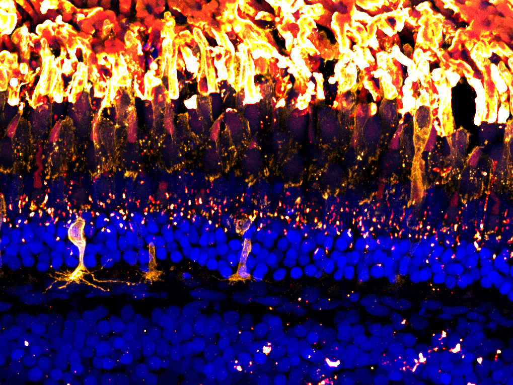
![Image by Natalia Jaroszynska](https://images.squarespace-cdn.com/content/v1/58065fb61b631b37ff3ce66a/1657560983493-TLSS4UR767W3NRYC28NU/DoctoralSchoolImageCompetition_adultretina_image11.png)
Image by Natalia Jaroszynska
![Actin cytoskeleton of Müller glia](https://images.squarespace-cdn.com/content/v1/58065fb61b631b37ff3ce66a/1675691195248-G09CUKYZQOPEYX7BMWUX/website2.jpg)
Actin cytoskeleton of Müller glia
Image by Natalia Jaroszynska
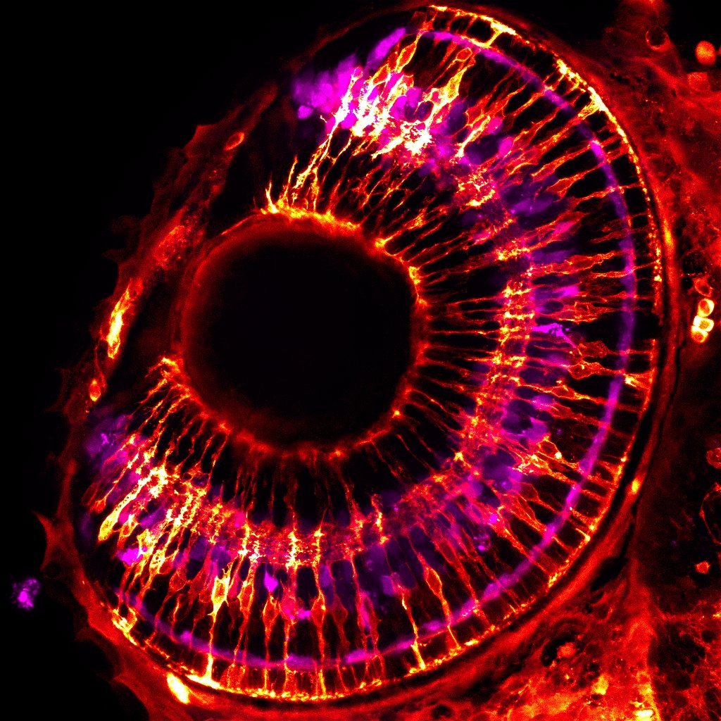
![3 day old zebrafish retina](https://images.squarespace-cdn.com/content/v1/58065fb61b631b37ff3ce66a/1657275827342-XUTVP654T9FBWQXN5PDT/DoctoralSchoolImageCompetition_DMSO_3dpf_image7.jpg)
3 day old zebrafish retina
Image by Natalia Jaroszynska
![Image by Gregory Patient Immunohistochemistry on a five days post fertilisation zebrafish retinal section, showing Müller glia, amacrine, retinal ganglion cells and F-Actin. The markers are: green – Glutamate Synthetase, cyan – HuC/D, magenta – Pha](https://images.squarespace-cdn.com/content/v1/58065fb61b631b37ff3ce66a/1678463596050-BVX3KKP2WX4943J9FH5P/image-asset.png)
Image by Gregory Patient
Immunohistochemistry on a five days post fertilisation zebrafish retinal section, showing Müller glia, amacrine, retinal ganglion cells and F-Actin. The markers are: green – Glutamate Synthetase, cyan – HuC/D, magenta – Phalloidin.
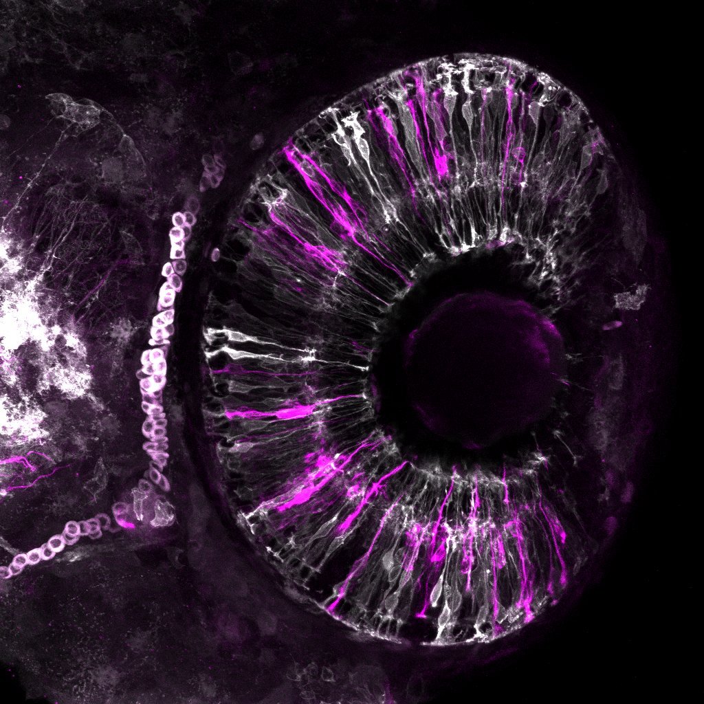
![Glial cytoskeleton](https://images.squarespace-cdn.com/content/v1/58065fb61b631b37ff3ce66a/1665150975646-AUPMKDKG22S0LJ9KYO4F/MAX_60HPF_EMTB-TdTomato_whole+eye_merge2.jpg)
Glial cytoskeleton
Image by Natalia Jaroszynska
Developing Müller glia (white) and their actin cytoskeleton (magenta) in a 2.5day old zebrafish retina.
![The inner plexiform layer](https://images.squarespace-cdn.com/content/v1/58065fb61b631b37ff3ce66a/1561391228963-IW0IBTGW5DNLZQJ668KP/Alternate4.jpg)
The inner plexiform layer
Image by Elisabeth Kugler
The inner plexiform layer is the major synaptic neurpil of the retina. In this layer Müller glia cell (multi-coloured) sends elaborate projections to contact and support neuronal synapses. Genotype Tg(Tp1:Venus) and false coloured in FIJI.
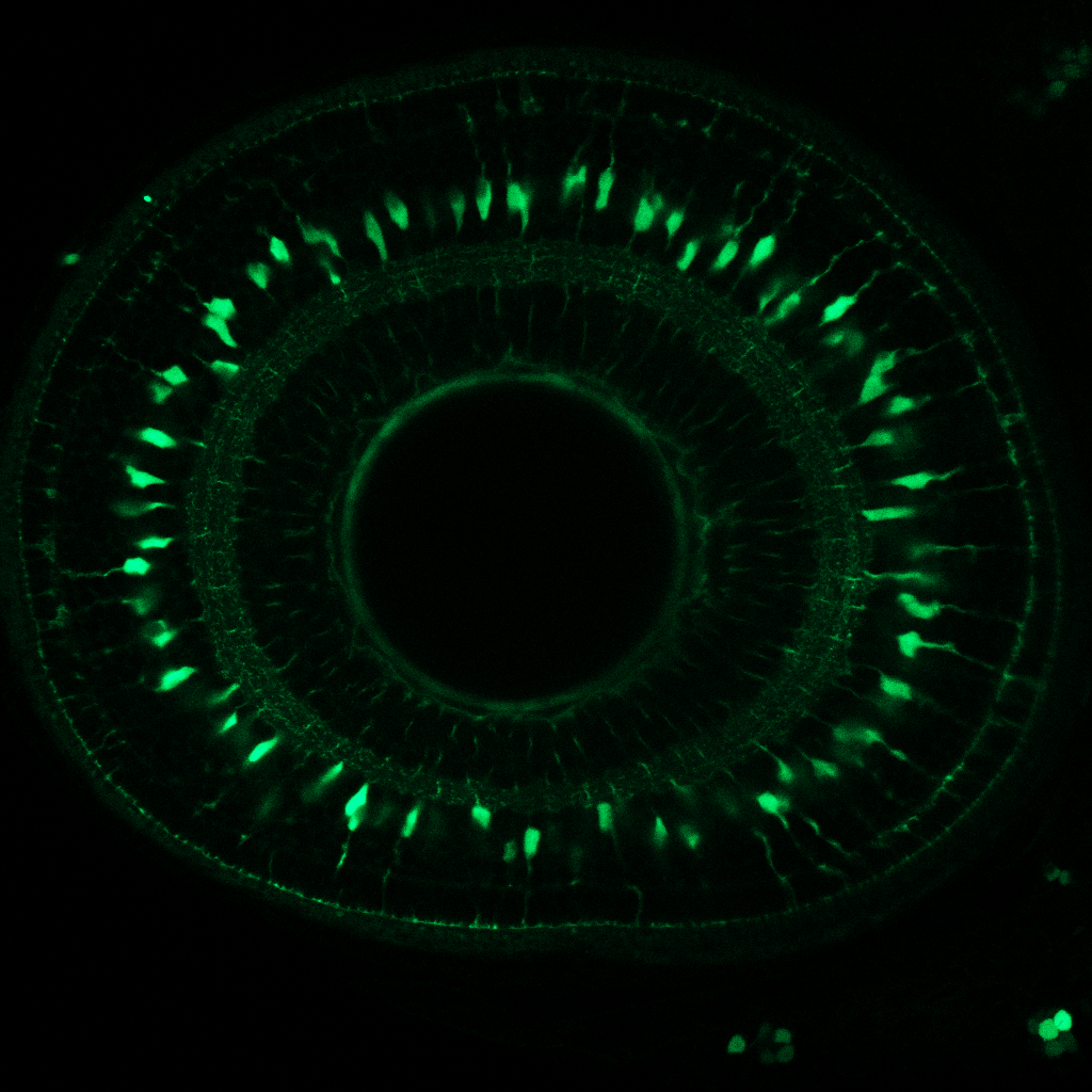
![Image by Manuela Lahne](https://images.squarespace-cdn.com/content/v1/58065fb61b631b37ff3ce66a/1677236448885-76DIKT8MWHQJ55FFW2CE/Manuela+1.png)
Image by Manuela Lahne
![Image by Elisabeth Kugler In the mature retina Müller glia cells “tile” to make an elaborate non-overlapping support network for neurons. Each Müller glia cell is morphologically specialised at each layer of the retina and will establish its’ own u](https://images.squarespace-cdn.com/content/v1/58065fb61b631b37ff3ce66a/1677518721397-MSNNMDVXXG75TQ090OYM/image-asset.png)
Image by Elisabeth Kugler
In the mature retina Müller glia cells “tile” to make an elaborate non-overlapping support network for neurons. Each Müller glia cell is morphologically specialised at each layer of the retina and will establish its’ own unique spatial domain during development. Optical section through the intact zebrafish retina with a Zeiss Airyscan confocal. Genotype Tg(Tp1:Venus) and false coloured in FIJI.
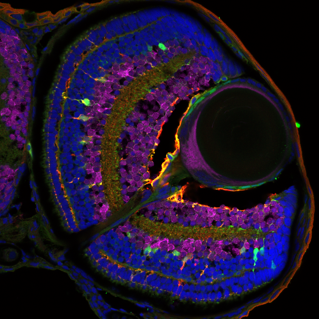
![Section of 96 hpf zebrafish retina Tg(tp1:Venus) anti-HuC/D in magenta, anti-GS in red and DAPI in blue](https://images.squarespace-cdn.com/content/v1/58065fb61b631b37ff3ce66a/1695992132332-80VA9NDA8MZDXUIV3QL4/96hpf+F1+GS+555+HucD+647+DAPI+Tp1Venus.png)
Section of 96 hpf zebrafish retina Tg(tp1:Venus) anti-HuC/D in magenta, anti-GS in red and DAPI in blue
Image by Aanandita Korthurkar
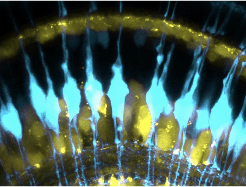
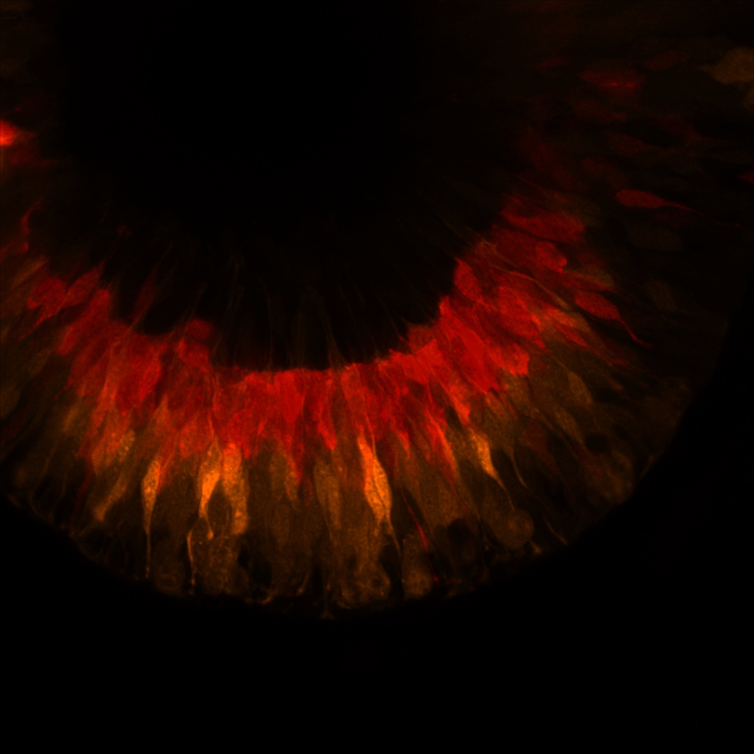
![Image by Ryan MacDonald The star of the show: In the retina the principal glial type are the Müller glia (blue). These cells are elaborately shaped to contact neurons (yellow) and provide them with support so they function properly. I want to know](https://images.squarespace-cdn.com/content/v1/58065fb61b631b37ff3ce66a/1677531626494-BCLLXJRDY495ANN2R89M/Screenshot+2023-02-27+at+20.56.13.png)
Image by Ryan MacDonald
The star of the show: In the retina the principal glial type are the Müller glia (blue). These cells are elaborately shaped to contact neurons (yellow) and provide them with support so they function properly. I want to know how these glial cells get their shape in development. Optical section through the intact zebrafish retina with a Zeiss airyscan confocal. Genotype Tg(Tp1:Venus;ptf1a:dsRED).
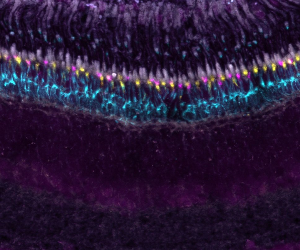
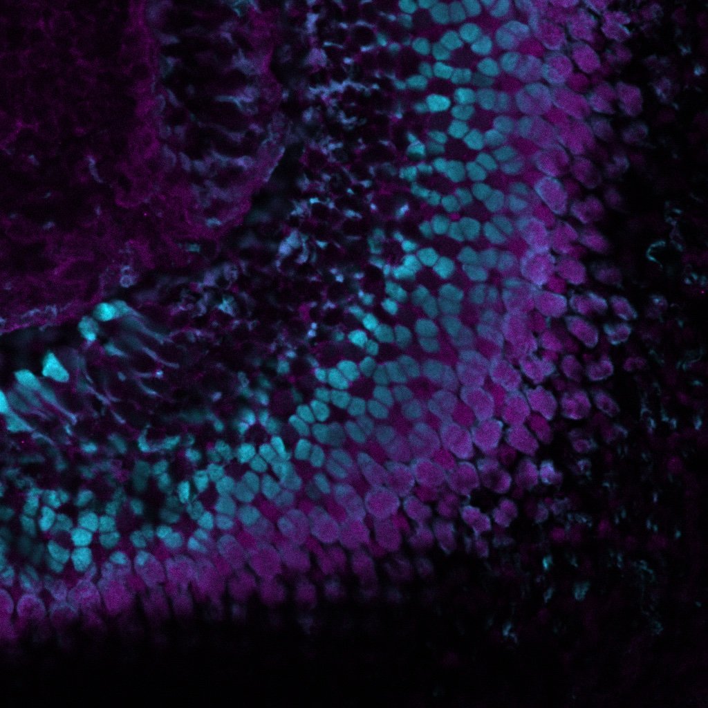
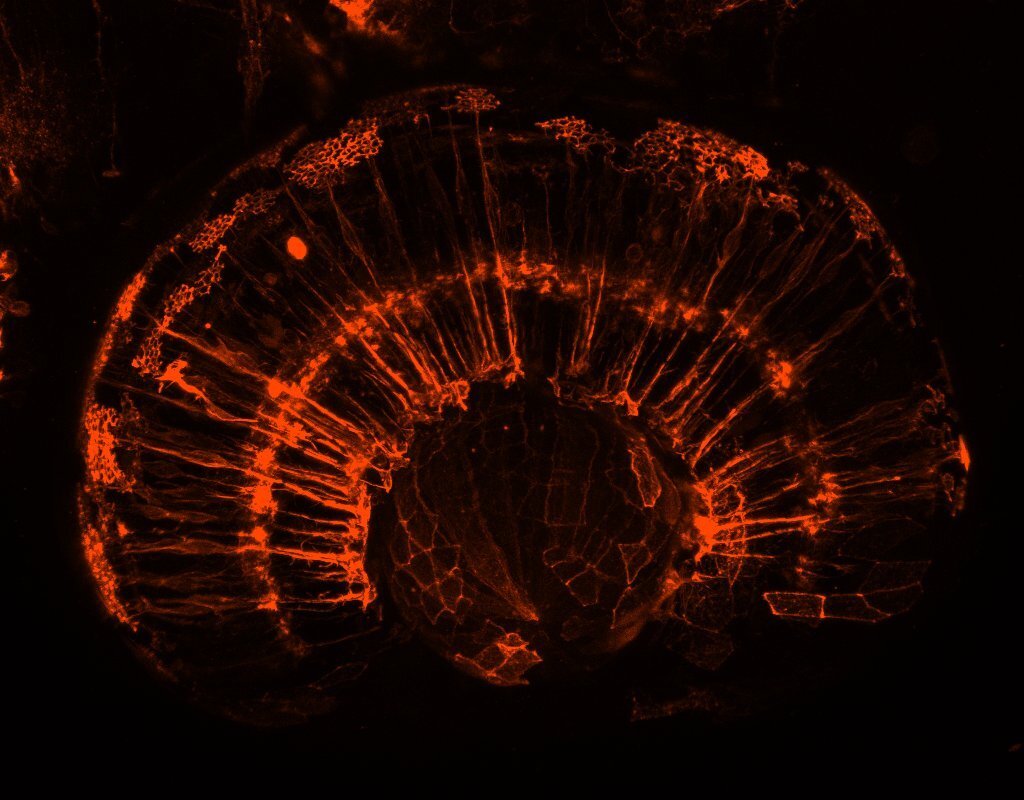
![HCR pRs killifish.jpg](https://images.squarespace-cdn.com/content/v1/58065fb61b631b37ff3ce66a/1695721714130-L1N7R9CU9R1SMIQDRPE0/HCR+pRs+killifish.jpg)
HCR pRs killifish.jpg
Image by Nicole Noel
In situ hybridization chain reaction on killifish retina. Showing photoreceptors.
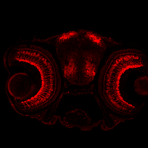
![Section of 5 dpf zebrafish retina anti-PKCb](https://images.squarespace-cdn.com/content/v1/58065fb61b631b37ff3ce66a/1695993661190-KBI8CK855TWESTD11PEG/new_aanandita_pkcB+546+20X+5dpf+WT+NEW.png)
Section of 5 dpf zebrafish retina anti-PKCb
Image by Aanandita Korthurkar
![Image by Xhuljana Durmishi](https://images.squarespace-cdn.com/content/v1/58065fb61b631b37ff3ce66a/1658414247554-3D5DOU89X4923F1LC68Q/1-72hpf_TP1eGFP-CAAX.jpg)
![Section of 96 hpf zerbafish retina Tg(tp1:Venus) anti-Ca1 in green, anti-zrf1 in red and DAPI in blue](https://images.squarespace-cdn.com/content/v1/58065fb61b631b37ff3ce66a/1695991078215-F4H9GQWI0TD3HUS3YOQ0/96hpf+F1+Carbonic+anhydrase+488+zrf1-550+Tp1Venus-1.png)
![By Xhuljana Durmishi](https://images.squarespace-cdn.com/content/v1/58065fb61b631b37ff3ce66a/1658414266419-BD37Q5ZEF86KFUGTN59T/4-48hpf_vsx1-ptf1a.jpg)
![Image by Gregory Patient A three round IBEX on a five days post fertilisation zebrafish retinal section, showing nuclei, different photoreceptor components, Müller glia, microglia, amacrine and retinal ganglion cells. The markers are: green – Gluta](https://images.squarespace-cdn.com/content/v1/58065fb61b631b37ff3ce66a/1678463460744-ENHF9T27QZTHAIXBI6LE/image-asset.png)
![Image by Manuela Lahne](https://images.squarespace-cdn.com/content/v1/58065fb61b631b37ff3ce66a/1674596371034-L761XDRK4UYTE6ZL1LOY/Manuela+3.png)
![Section cell body.jpg](https://images.squarespace-cdn.com/content/v1/58065fb61b631b37ff3ce66a/1695721946747-6K1IGZPWDZPZN974LIQV/Section+cell+body.jpg)
![Glutamine synthetase](https://images.squarespace-cdn.com/content/v1/58065fb61b631b37ff3ce66a/1561391286298-9Y40FUHBW8YWJ54NSJI2/Publications.jpg)
![Image by Ryan MacDonald](https://images.squarespace-cdn.com/content/v1/58065fb61b631b37ff3ce66a/1671201881984-FYV17ZE0340O6341ZL4X/MullerGlia-AC.png)
![By Xhuljana Durmishi](https://images.squarespace-cdn.com/content/v1/58065fb61b631b37ff3ce66a/1658414275310-S0D3Y7K30OY9SQ5ZZB6L/6-48hpf_vsx1-ptf1a.jpg)
![Image by Elisabeth Kugler False-colour illustration of zebrafish retina neurons (blue) and glia progenitors (yellow) at 48 hours post fertilization, acquired with Zeiss AiryScan microscopy. Image processing was conducted with Fiji software.](https://images.squarespace-cdn.com/content/v1/58065fb61b631b37ff3ce66a/1612265628387-HIHJYVKPCZCMGNJKGWAH/Composite.jpg)
![Tg(gfap:gfp)](https://images.squarespace-cdn.com/content/v1/58065fb61b631b37ff3ce66a/1561557824533-DFUWXIV5D01XPDFUX14M/Picture+2+%281%29.jpg)
![Müller glia and retinal neurons](https://images.squarespace-cdn.com/content/v1/58065fb61b631b37ff3ce66a/1657562192266-IN3CCEW12ND3X4O0N2PY/website+pic+2.png)
![Glial cells support healthy neurons](https://images.squarespace-cdn.com/content/v1/58065fb61b631b37ff3ce66a/1561391211363-TXPDAGTWDOGUEALGIOD9/Alternate+2.jpg)
![The retina connects to the visual centres in the brain](https://images.squarespace-cdn.com/content/v1/58065fb61b631b37ff3ce66a/1561391215257-VZSYUQAP98ZUCO2IE2LV/Alternate1.jpg)
![202305 zpr-1-magenta_DAPI-cyan_12wo-killifish.jpg](https://images.squarespace-cdn.com/content/v1/58065fb61b631b37ff3ce66a/1695721684502-UTSOCJYZSI1GTN0B0RYY/202305+zpr-1-magenta_DAPI-cyan_12wo-killifish.jpg)
![Semi-super resolution reconstruction of Müller glia cells](https://images.squarespace-cdn.com/content/v1/58065fb61b631b37ff3ce66a/1561557298181-FJAGIYVZH1RLU8GZQ3SO/Alternate5.jpg)
![HCR in a 4-day old zebrafish transgenic (tp1:Venus) retina, showing Vsx1 (magenta) mRNA](https://images.squarespace-cdn.com/content/v1/58065fb61b631b37ff3ce66a/1657561390038-B5R7WW6HBB1EAPTL4IXF/website+pic.png)
![Muller glia in the zebrafish retina](https://images.squarespace-cdn.com/content/v1/58065fb61b631b37ff3ce66a/1612265619481-8JIBTDTG0ZJCA1NT6VN6/001.png)
![Image by Xhuljana Durmishi](https://images.squarespace-cdn.com/content/v1/58065fb61b631b37ff3ce66a/1658414252874-JBCUENJFAIPNBKW5IRFF/2-72hpf_vsx1-ptf1a.jpg)
![zpr-3 killifish.jpg](https://images.squarespace-cdn.com/content/v1/58065fb61b631b37ff3ce66a/1695728855420-70L5SDFA6EX2VBBAFE1T/zpr-3+killifish.jpg)
![Image by Elisabeth Kugler Image shows multi-colour false segmentation of the zebrafish retina at 3dpf. Image processing was conducted with Fiji software.](https://images.squarespace-cdn.com/content/v1/58065fb61b631b37ff3ce66a/1612265630739-F36DBAN7I0ZYMAUFY2V2/interactive+watershed-Z.jpg)
![Muller glia connections.](https://images.squarespace-cdn.com/content/v1/58065fb61b631b37ff3ce66a/1612265635300-Y32MCCZL3FNKJYU2ZW7J/MAX_z-reduced_60hpf_ath5gapGFP_cslmCherry_e1-01-Airyscan+Processing-482.jpg)
![Between the eyes](https://images.squarespace-cdn.com/content/v1/58065fb61b631b37ff3ce66a/1657560940377-A9BHBUCWX06B35D8JEFH/Snap-542.jpg)
![Image by Gregory Patient A multiplex of four rabbit antibodies on the same five days post fertilisation zebrafish retinal section using micro-conjugation kits. Photoreceptors shown in green by GNAT2, Müller glia cells in magenta with RLBP1 and syna](https://images.squarespace-cdn.com/content/v1/58065fb61b631b37ff3ce66a/1678463653393-UHJPQ071IR2QZZ4M0LY1/image-asset.png)
![Muller glia support synapses](https://images.squarespace-cdn.com/content/v1/58065fb61b631b37ff3ce66a/1612265979583-HYXTOUS7J603A5I0GHQF/Screenshot+2020-10-30+at+11.32.15.png)
![Image by Manuela Lahne](https://images.squarespace-cdn.com/content/v1/58065fb61b631b37ff3ce66a/1674596363691-KGMAYAPGF71YZNWQVPOJ/manuela+2.png)
![Image by Elisabeth Kugler](https://images.squarespace-cdn.com/content/v1/58065fb61b631b37ff3ce66a/1629817340500-SQ08SU87E27KV9G5AHUL/FocalPlane_01.jpg)
![Image by Elisabeth Kugler](https://images.squarespace-cdn.com/content/v1/58065fb61b631b37ff3ce66a/1629817346649-0Z60N1A3XI7SSZ19BNM5/MAX_colored_60hpf.jpg)
![Image by Elisabeth Kugler](https://images.squarespace-cdn.com/content/v1/58065fb61b631b37ff3ce66a/1629817381551-6OTMM29WS5OKWP5WK1XG/Alicia.jpg)
![Image by Elisabeth Kugler](https://images.squarespace-cdn.com/content/v1/58065fb61b631b37ff3ce66a/1655046777737-NYHR75LB16PUTXCCOSCU/FU4-k2XX0AA4tU-.jpg)
![HCR in 5 dpf wildtype zebrafish retina, showing cyp26a1 mRNA](https://images.squarespace-cdn.com/content/v1/58065fb61b631b37ff3ce66a/1695991785527-S57QB3RQA4HCJQSTLGL0/MAX_17-5-23+Cdn+2+A-A+cyp26a1+B1+488+glula+b3+546-01.czi+-+17-5-23+Cdn+2+A-A+cyp26a1+B1+488+glula+b3+546-01.czi+%233-1.png)
![Killifish retina](https://images.squarespace-cdn.com/content/v1/58065fb61b631b37ff3ce66a/1669983637847-3EV16ZWWSPRPXQM3P7P8/M_8+bits_Scale+bar_11.11.21+female+Killifish+retina+2+months+old+DAPI+GS+647+GFAP+488+40x+objective+speed+5s_Substack+%281-10%29.jpg)
![Image by Isabel Bravo](https://images.squarespace-cdn.com/content/v1/58065fb61b631b37ff3ce66a/1671201882221-028LGFLLYFAZGE7CR312/MullerGlia.png)
![zpr-1 killifish 2.jpg](https://images.squarespace-cdn.com/content/v1/58065fb61b631b37ff3ce66a/1695728855123-VOUGISVNNA4JMD2KG0PI/zpr-1+killifish+2.jpg)
![By Xhuljana Durmishi](https://images.squarespace-cdn.com/content/v1/58065fb61b631b37ff3ce66a/1658414255606-CXU3MQPZWBBZADGF2I9U/3-72hpf_TP1Venus_immuno.jpg)
![By Xhuljana Durmishi](https://images.squarespace-cdn.com/content/v1/58065fb61b631b37ff3ce66a/1658414269755-XAE49TMDUDK2RX9GMMWH/5-72hpf_TP1-krdl.jpg)
![Actin cytoskeleton of Müller glia](https://images.squarespace-cdn.com/content/v1/58065fb61b631b37ff3ce66a/1675691195090-G8V8WHP0IJD68J7D29HC/MG+actin+cytoskeleton.png)
![Image by Natalia Jaroszynska](https://images.squarespace-cdn.com/content/v1/58065fb61b631b37ff3ce66a/1657560983493-TLSS4UR767W3NRYC28NU/DoctoralSchoolImageCompetition_adultretina_image11.png)
![Actin cytoskeleton of Müller glia](https://images.squarespace-cdn.com/content/v1/58065fb61b631b37ff3ce66a/1675691195248-G09CUKYZQOPEYX7BMWUX/website2.jpg)
![3 day old zebrafish retina](https://images.squarespace-cdn.com/content/v1/58065fb61b631b37ff3ce66a/1657275827342-XUTVP654T9FBWQXN5PDT/DoctoralSchoolImageCompetition_DMSO_3dpf_image7.jpg)
![Image by Gregory Patient Immunohistochemistry on a five days post fertilisation zebrafish retinal section, showing Müller glia, amacrine, retinal ganglion cells and F-Actin. The markers are: green – Glutamate Synthetase, cyan – HuC/D, magenta – Pha](https://images.squarespace-cdn.com/content/v1/58065fb61b631b37ff3ce66a/1678463596050-BVX3KKP2WX4943J9FH5P/image-asset.png)
![Glial cytoskeleton](https://images.squarespace-cdn.com/content/v1/58065fb61b631b37ff3ce66a/1665150975646-AUPMKDKG22S0LJ9KYO4F/MAX_60HPF_EMTB-TdTomato_whole+eye_merge2.jpg)
![The inner plexiform layer](https://images.squarespace-cdn.com/content/v1/58065fb61b631b37ff3ce66a/1561391228963-IW0IBTGW5DNLZQJ668KP/Alternate4.jpg)
![Image by Manuela Lahne](https://images.squarespace-cdn.com/content/v1/58065fb61b631b37ff3ce66a/1677236448885-76DIKT8MWHQJ55FFW2CE/Manuela+1.png)
![Image by Elisabeth Kugler In the mature retina Müller glia cells “tile” to make an elaborate non-overlapping support network for neurons. Each Müller glia cell is morphologically specialised at each layer of the retina and will establish its’ own u](https://images.squarespace-cdn.com/content/v1/58065fb61b631b37ff3ce66a/1677518721397-MSNNMDVXXG75TQ090OYM/image-asset.png)
![Section of 96 hpf zebrafish retina Tg(tp1:Venus) anti-HuC/D in magenta, anti-GS in red and DAPI in blue](https://images.squarespace-cdn.com/content/v1/58065fb61b631b37ff3ce66a/1695992132332-80VA9NDA8MZDXUIV3QL4/96hpf+F1+GS+555+HucD+647+DAPI+Tp1Venus.png)
![Image by Ryan MacDonald The star of the show: In the retina the principal glial type are the Müller glia (blue). These cells are elaborately shaped to contact neurons (yellow) and provide them with support so they function properly. I want to know](https://images.squarespace-cdn.com/content/v1/58065fb61b631b37ff3ce66a/1677531626494-BCLLXJRDY495ANN2R89M/Screenshot+2023-02-27+at+20.56.13.png)
![HCR pRs killifish.jpg](https://images.squarespace-cdn.com/content/v1/58065fb61b631b37ff3ce66a/1695721714130-L1N7R9CU9R1SMIQDRPE0/HCR+pRs+killifish.jpg)
![Section of 5 dpf zebrafish retina anti-PKCb](https://images.squarespace-cdn.com/content/v1/58065fb61b631b37ff3ce66a/1695993661190-KBI8CK855TWESTD11PEG/new_aanandita_pkcB+546+20X+5dpf+WT+NEW.png)