
Gaia Gestri and Steve Wilson
Original paper reference:
Six3 functions in anterior neural plate specification by promoting cell proliferation and inhibiting Bmp4 expression
During development of the embryo, cells make decisions about the tissues they will contribute to and how they will differentiate. The outer layer of the embryo is called ectoderm and cells in this layer make the nervous system (brain and spinal cord) and the skin as well as migratory cells called neural crest. How do the ectoderm cells know which of these tissues to form? Within cells there are genes that encode proteins called transcription factors that act as molecular switches turning on different developmental programmes that control cell identity. In this study, we explored how one such transcription factor, called Six3, promotes the ability of cells to form eyes and brain.
Six3 is sometimes called a 'master regulator' for eye and forebrain development in that it is necessary and sufficient for the eyes and brain to form. What this means is that in gain of function experiments (in which the embryos express more Six3 protein), embryos show brain enlargement and ectopic eye structures (see the arrow pointing to 'the third eye' in Fig.1) while in loss of function experiments (where the Six3 protein is absent or not functioning) embryos show reduction of the most anterior part of the brain and no eyes (Fig. 1, right).
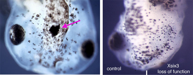
Figure 1. Pictures of the heads of frog tadpoles the black blobs are the forming eyes. In the tadpole on the left there is a small extra eye on top of the head when there is too much Six3 protein and the tadpole on the right is missing an eye due to lacking Six3 protein on one side of its brain.
So how might Six3 control brain and eye development? With this in mind, we asked when and how Six3 exerts it function taking advantage of both the frog, Xenopus laevis, and the zebrafish as model systems for our experiments.
The processes that regulate brain development start at the early neural plate stage, this is much earlier than the first morphological appearance of the eye and the brain. The neural plate is a sheet of cells that will form both the brain and the eyes and at first, it is continuous with a sheet of cells called the epidermis that will form the skin. It is the anterior part of the neural plate, ANP, (Fig. 2 red) that gives rise to the brain and eyes (Fig. 2).
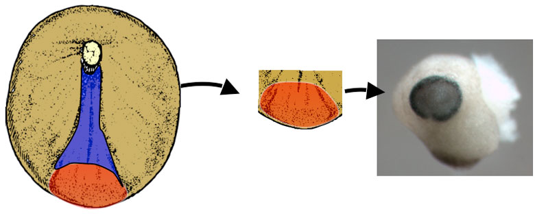
Figure 2. The schematic shows a very young frog embryo where the cells that are going to form the nervous system are coloured in red and blue. The red cells are those that will develop into the brain and eyes (right photo)
Given that the ANP has the capacity to form the brain and eyes, then the key genes that control eye and brain development must already be active in the ANP. These ANP genes must positively confer brain/eye identity as well making sure that the cells don't acquire the identity of the surrounding tissues such as the posterior neural plate (blue in Fig. 2), that will give rise to the spinal cord, and the surrounding epidermis that will give rise to the skin (brown in Fig 2). In our study we found that Six3 plays key roles in both specification and maintenance of the brain/eye promoting properties of the ANP.

Figure 3. Schematic and two photos of the anterior neural plate (blue in the schematic and white in the two photos. The neural plate is surrounded by epidermis (yellow in the schematic and blue in the photos). The little brown dots in the photos are individual cells that either have a lot of Six3 (middle), or lack Six3 (right)
One of the properties of the ANP is that it is more highly proliferative compared with the posterior neural plate and we found that Six3, expressed in this territory, promotes proliferation and represses the production of neurons.
The ANP is surrounded by the epidermis, (Fig. 3) that will later form the skin. The epidermis produces a signal, called Bmp, that has the ability to inhibit expression of ANP genes. We found that Six3 switches off Bmp expression (Fig. 3 middle) ensuring the maintenance of the ANP as a Bmp-free domain. This is really important given that if Six3 function is lost, Bmp signalling encroaches into the ANP (Fig. 3 right) and doing so, leads to embryos with severely reduced anterior brain and eyes and expanded epidermis (Fig. 1 right panel).
In humans, mutations in Six3 can lead to severe congenital abnormalities of the brain and eyes, and this study has helped to elucidate the function of this gene, one of the most important genes for brain and eye development.
This study was done as a collaboration between Guiseppina Barsacchi's lab in Pisa and Steve Wilson's lab here at UCL.