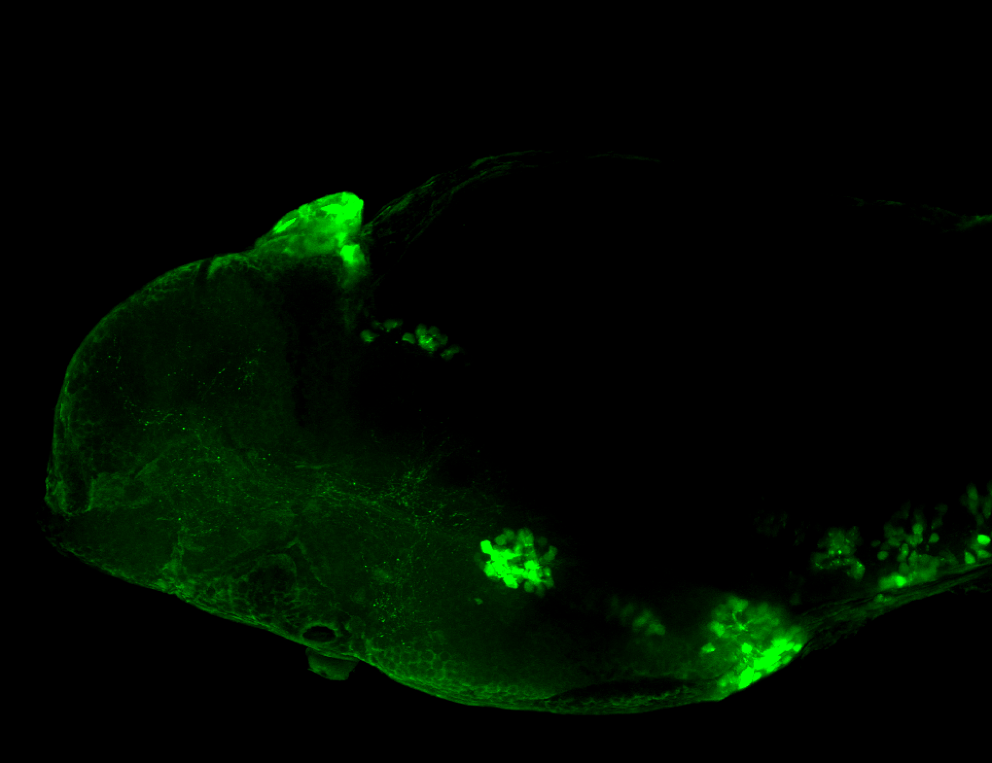
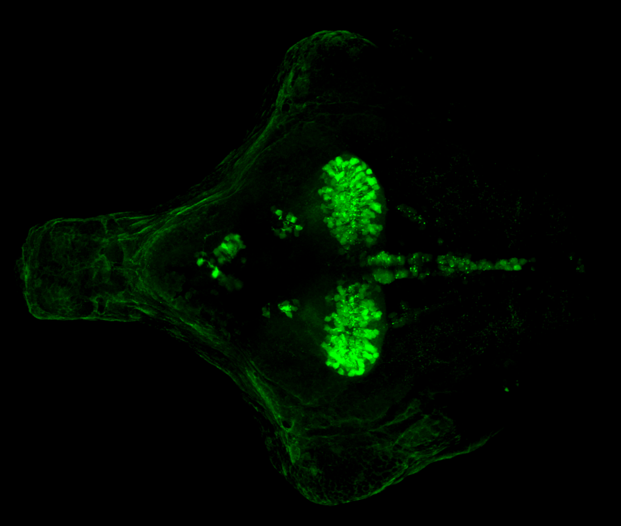
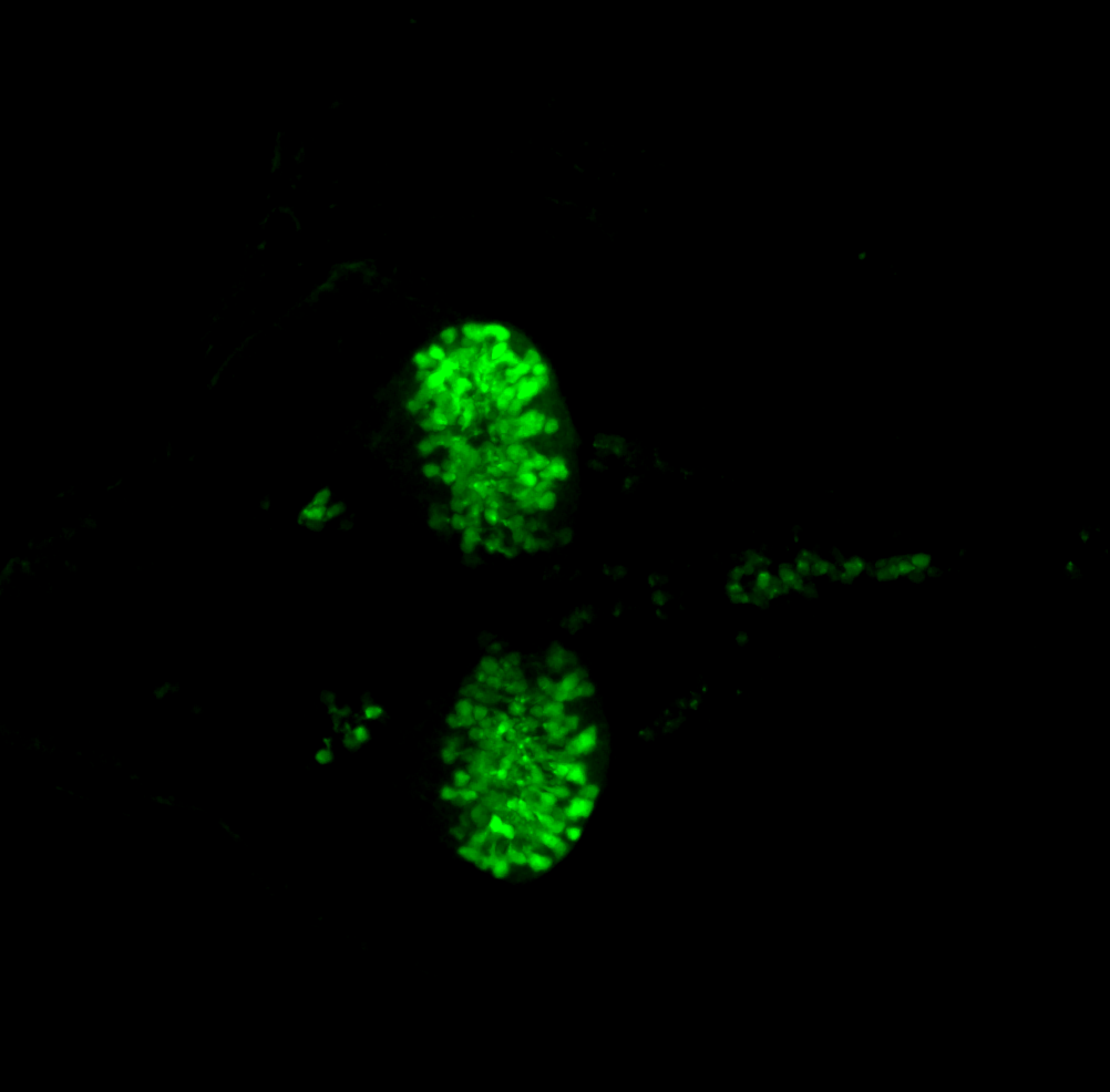
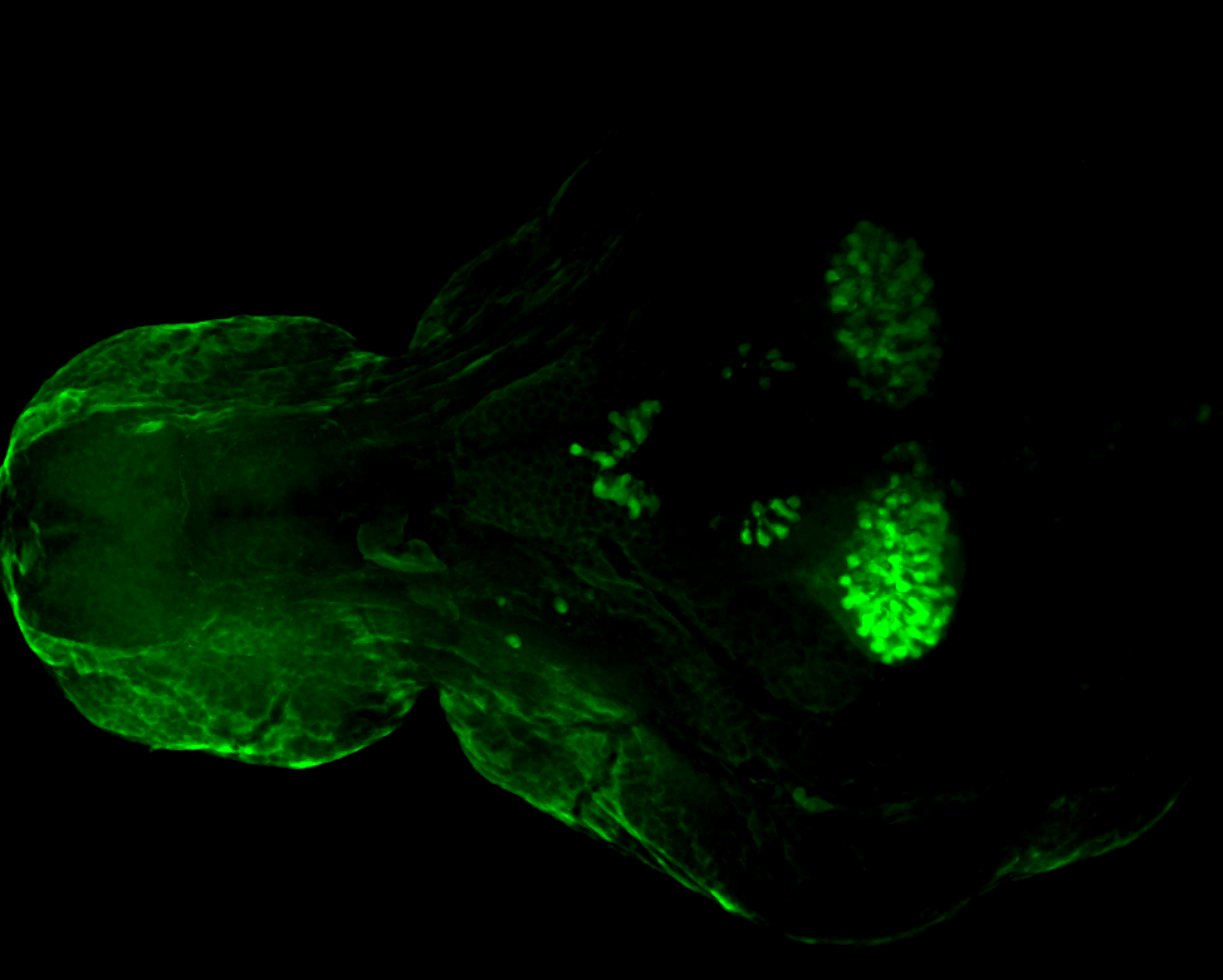
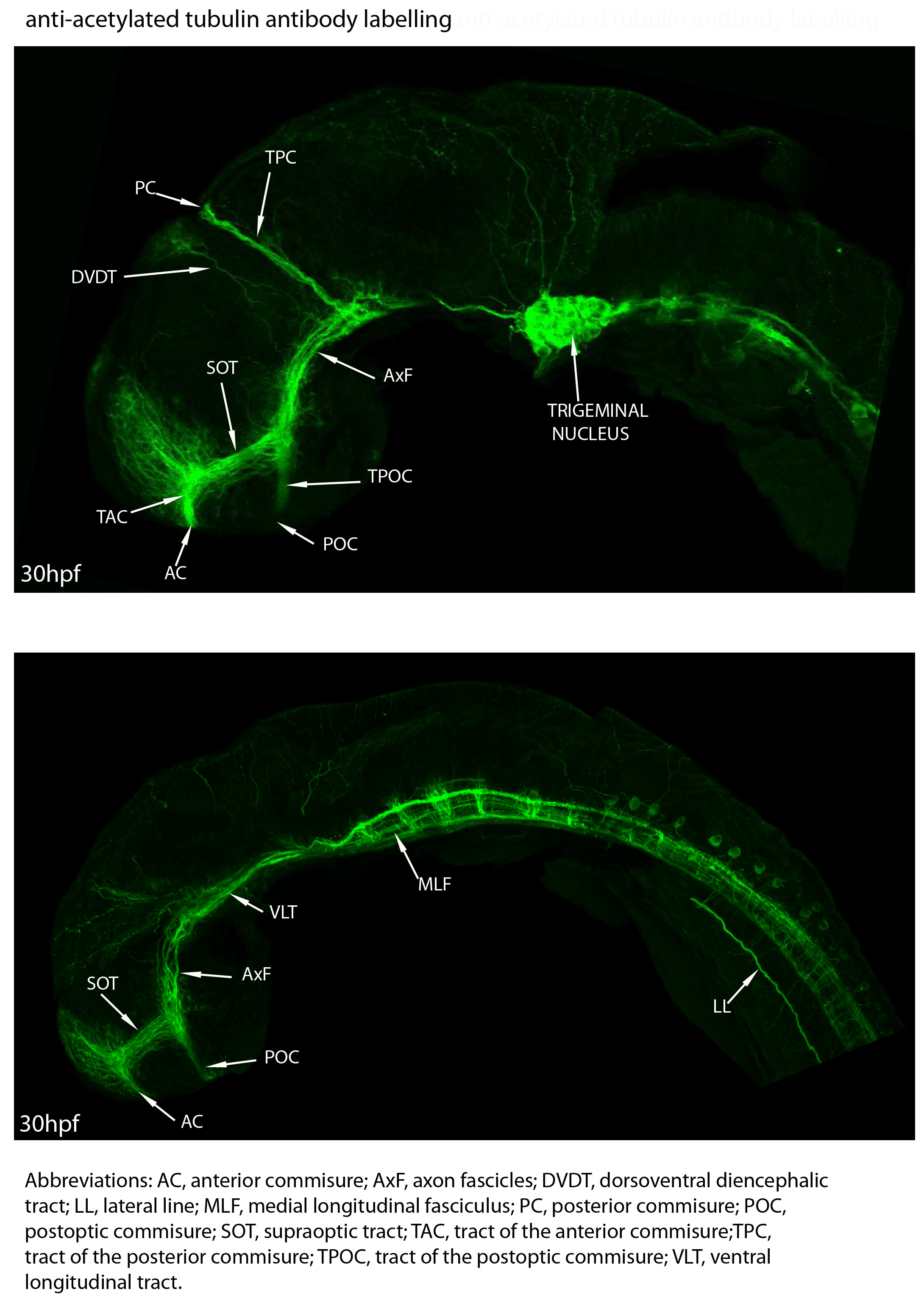
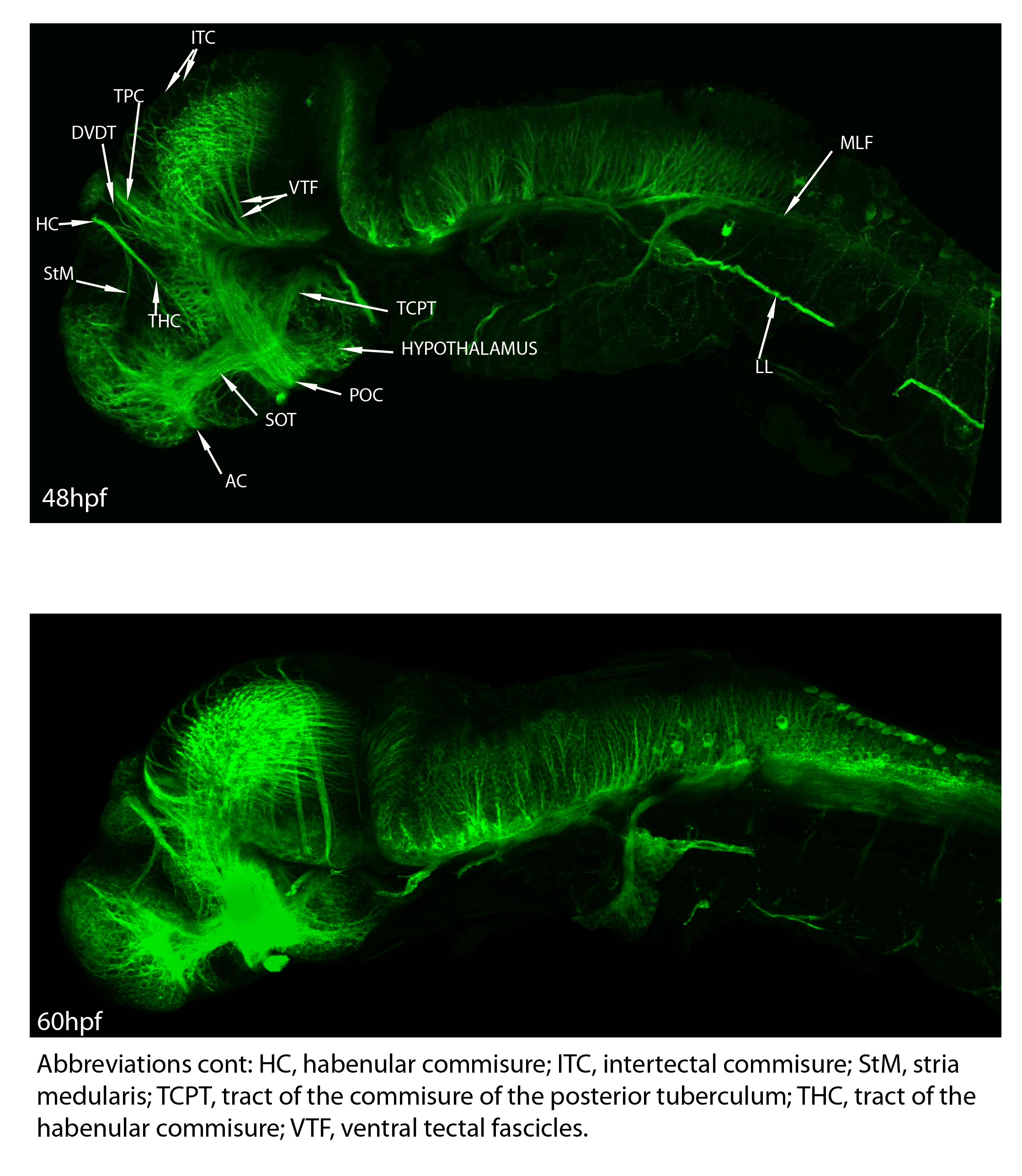
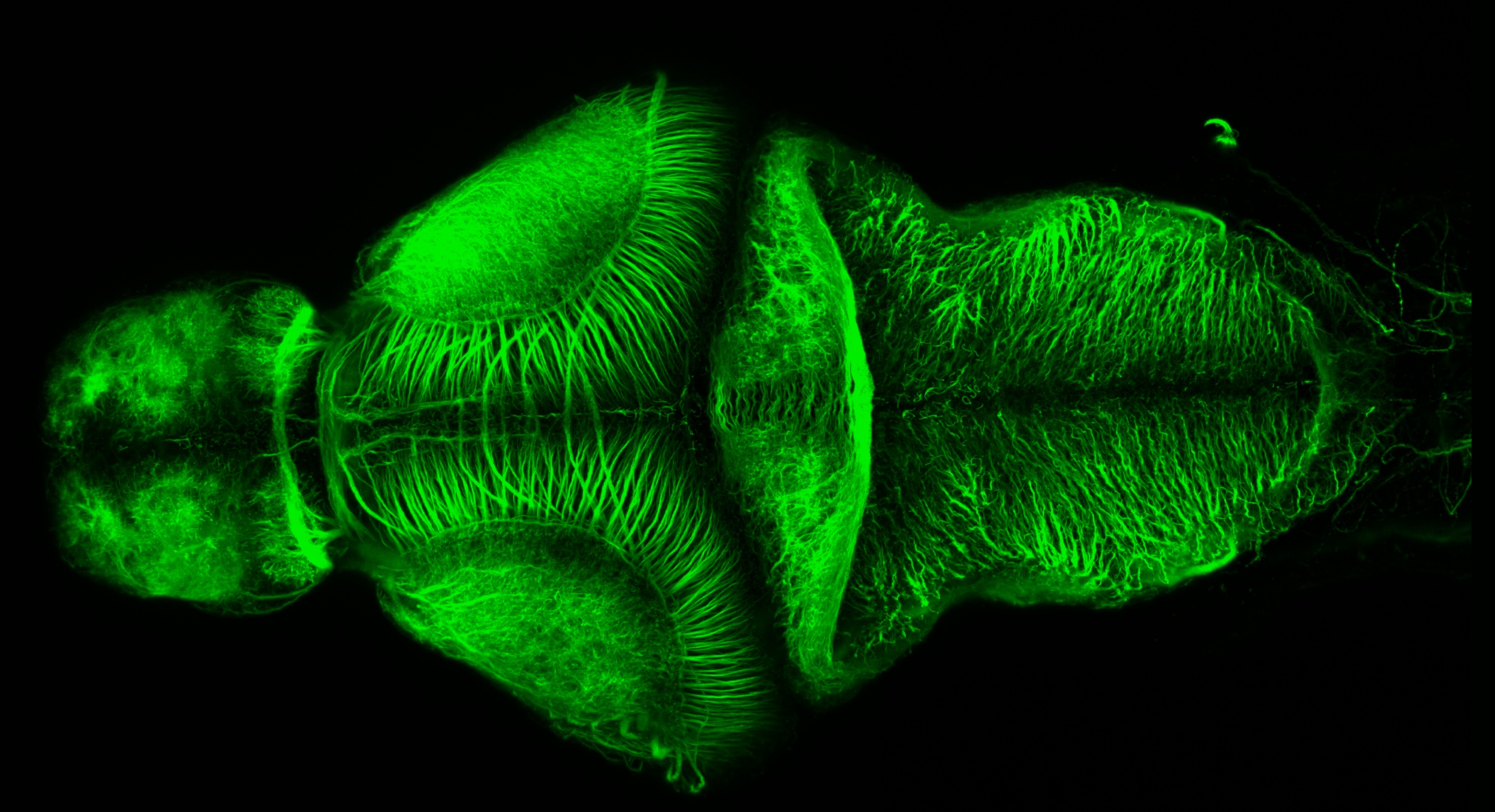
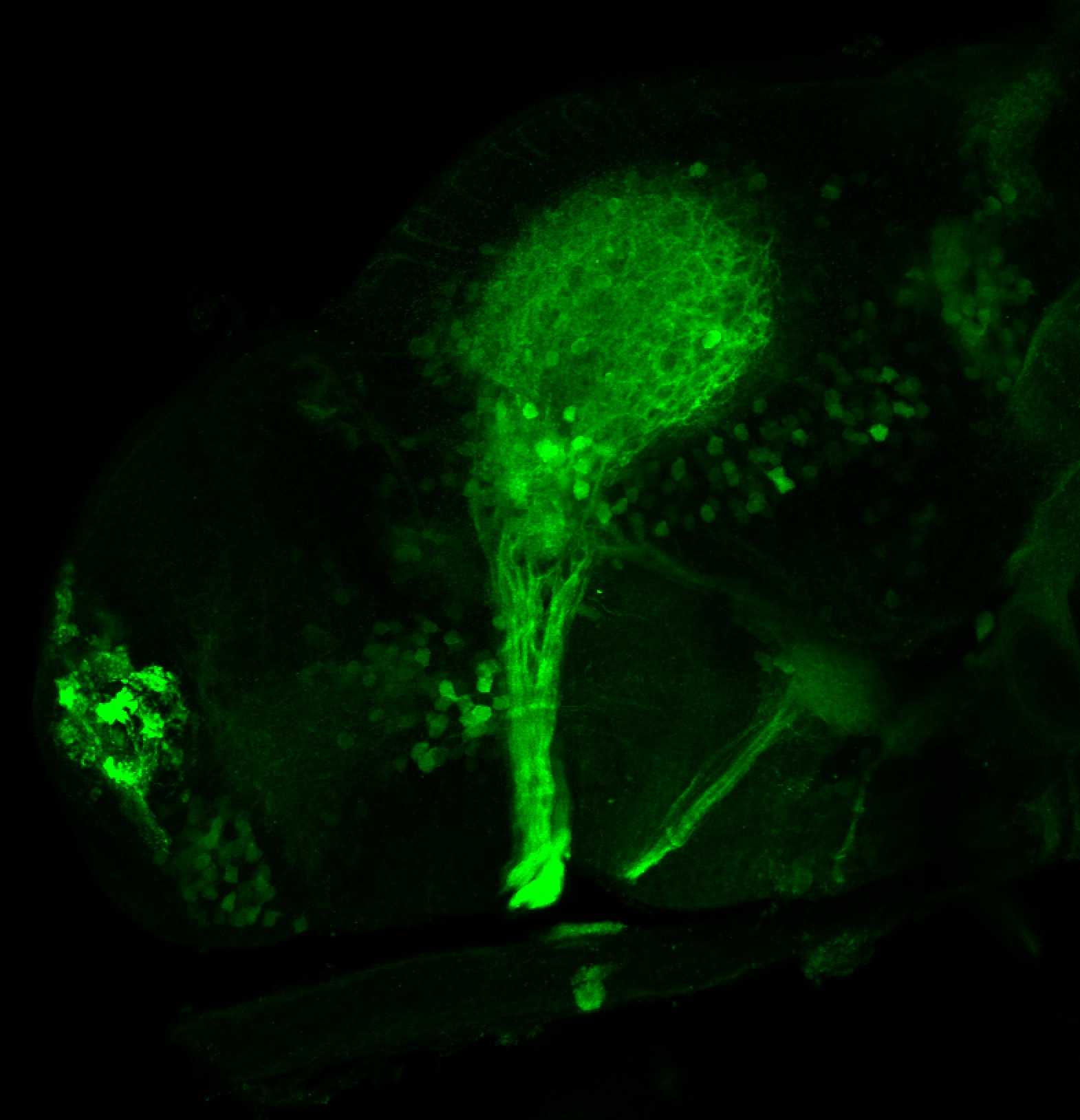
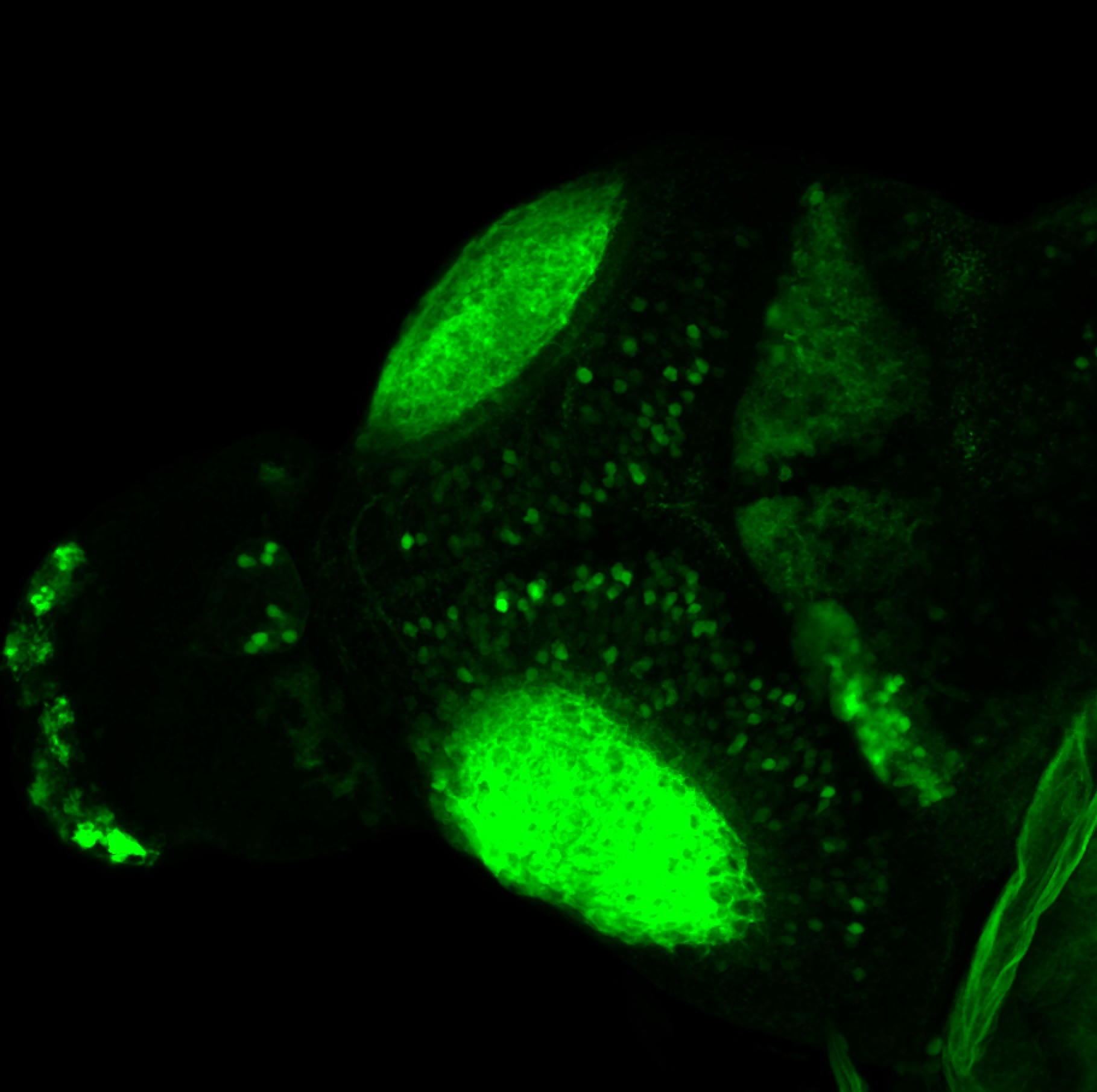
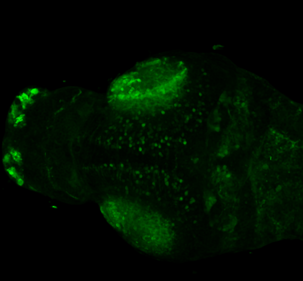
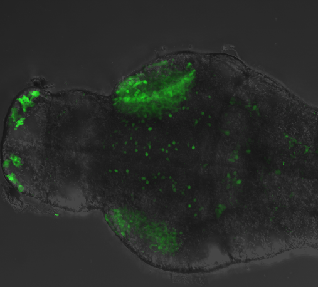
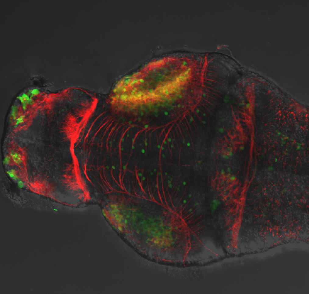
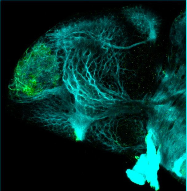
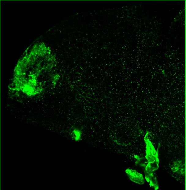
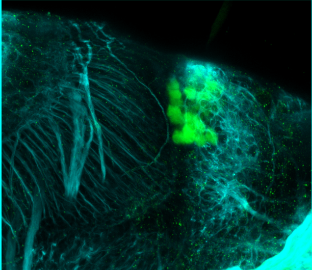
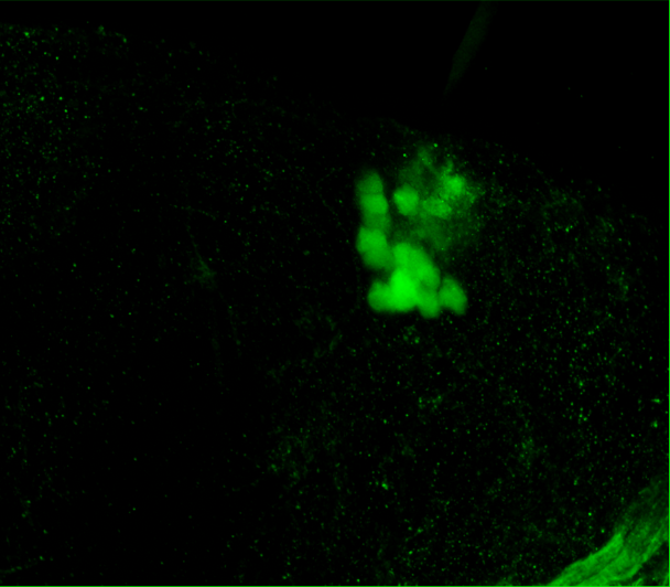
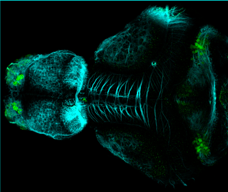
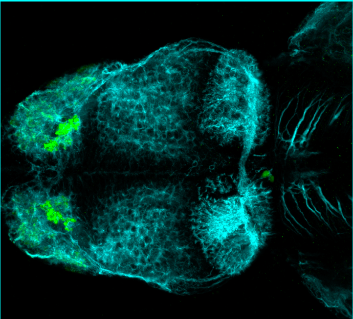
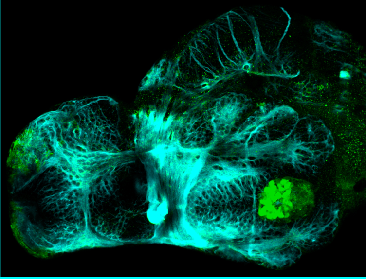
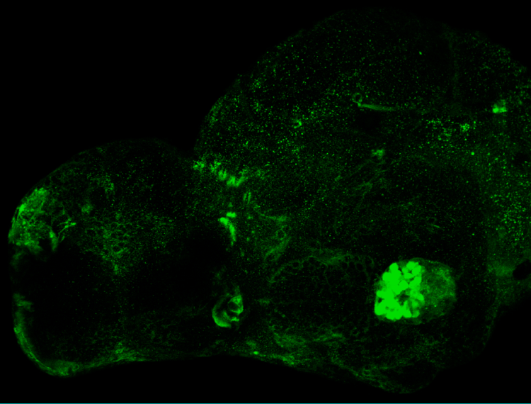
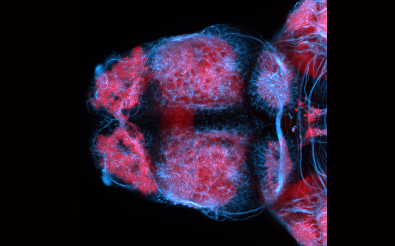
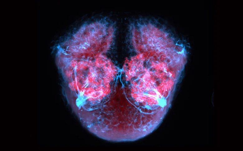
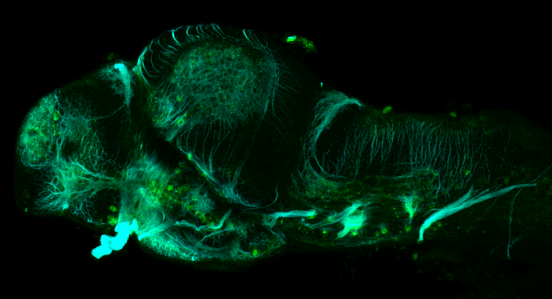
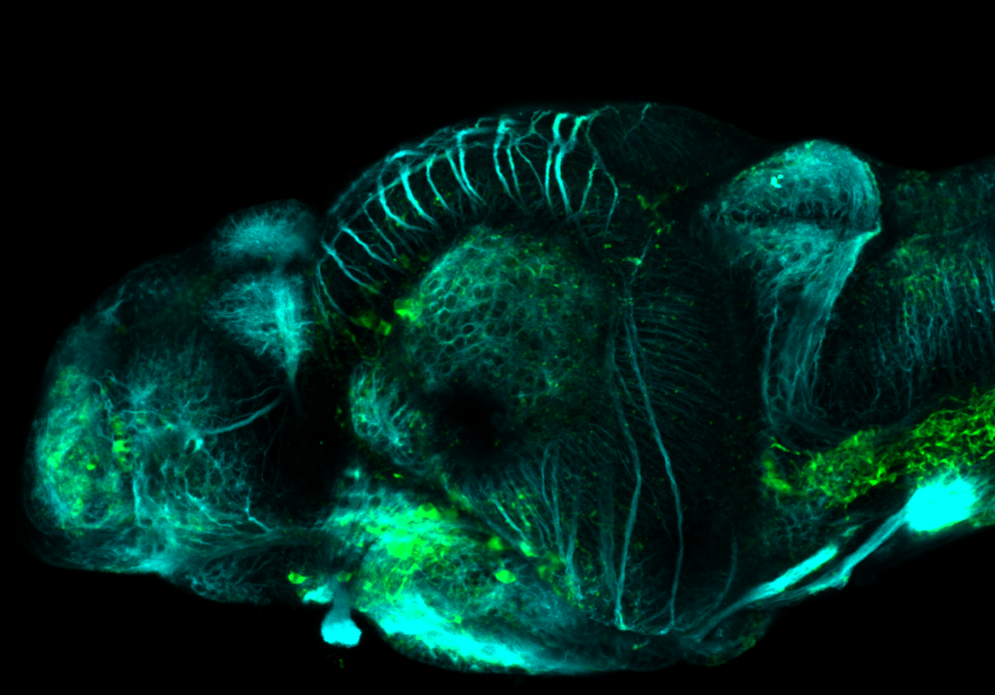
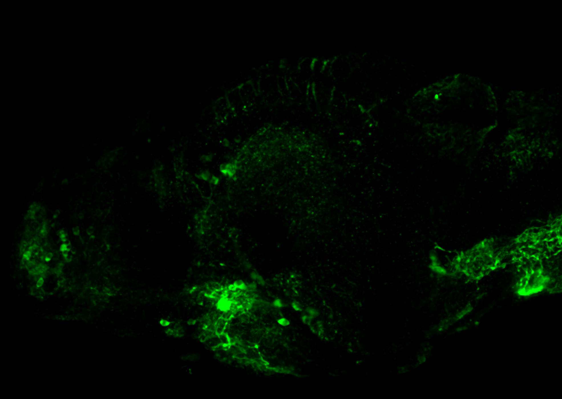
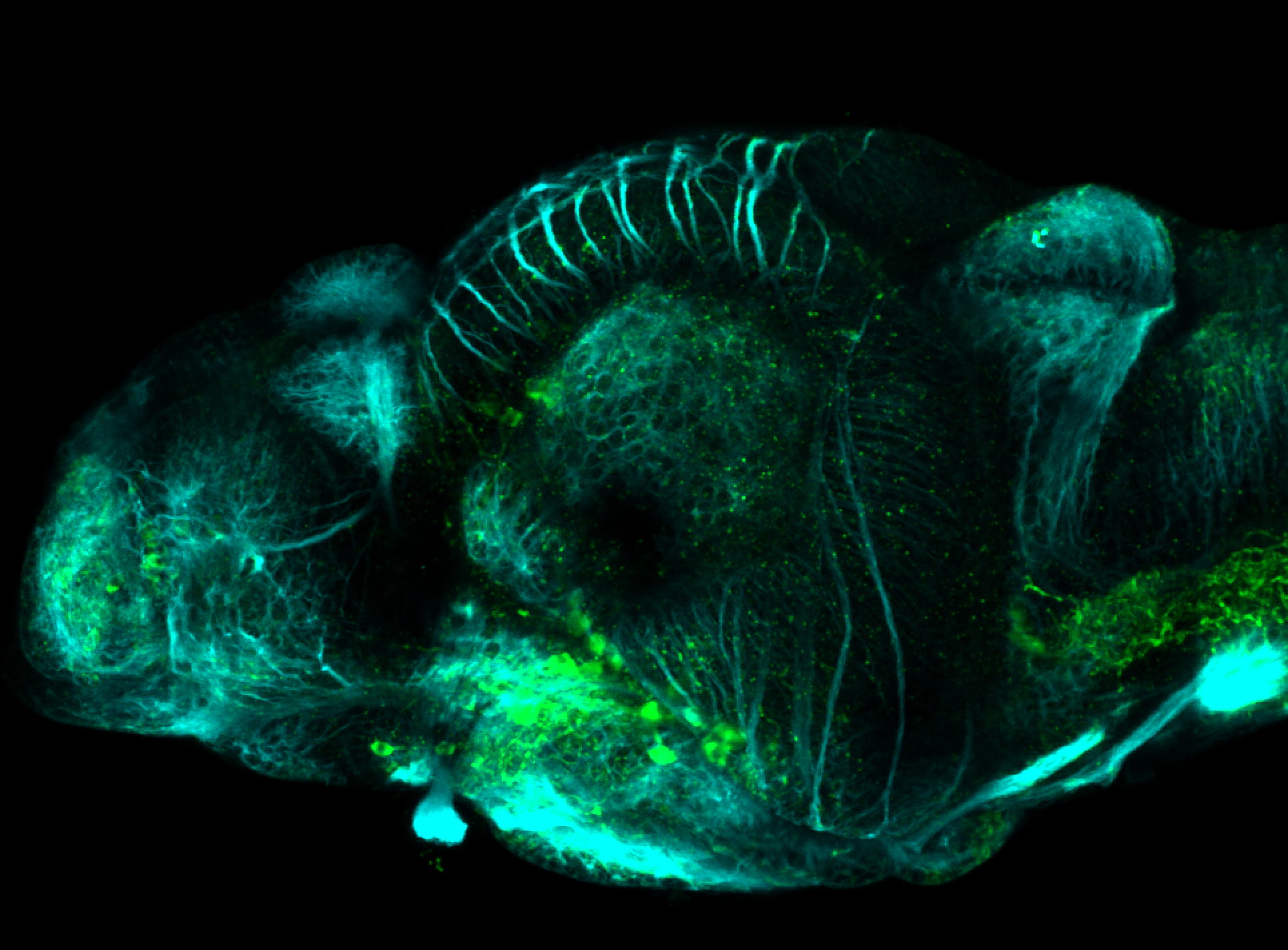
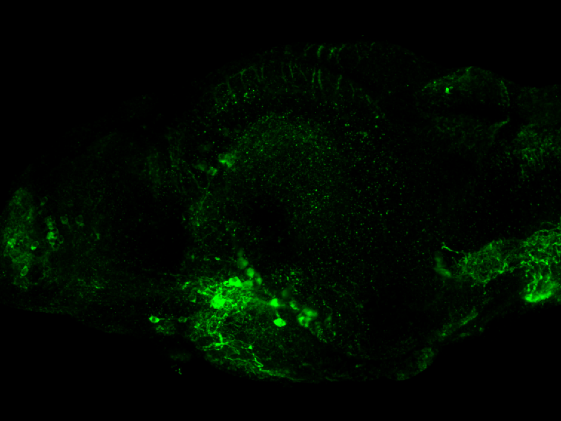
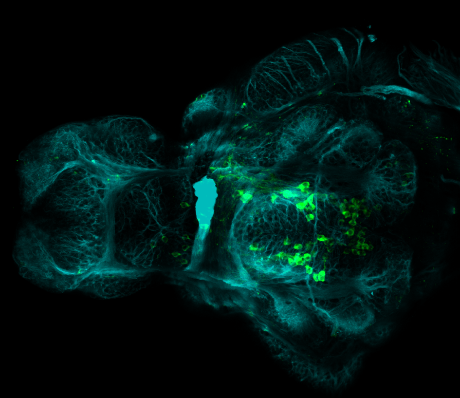
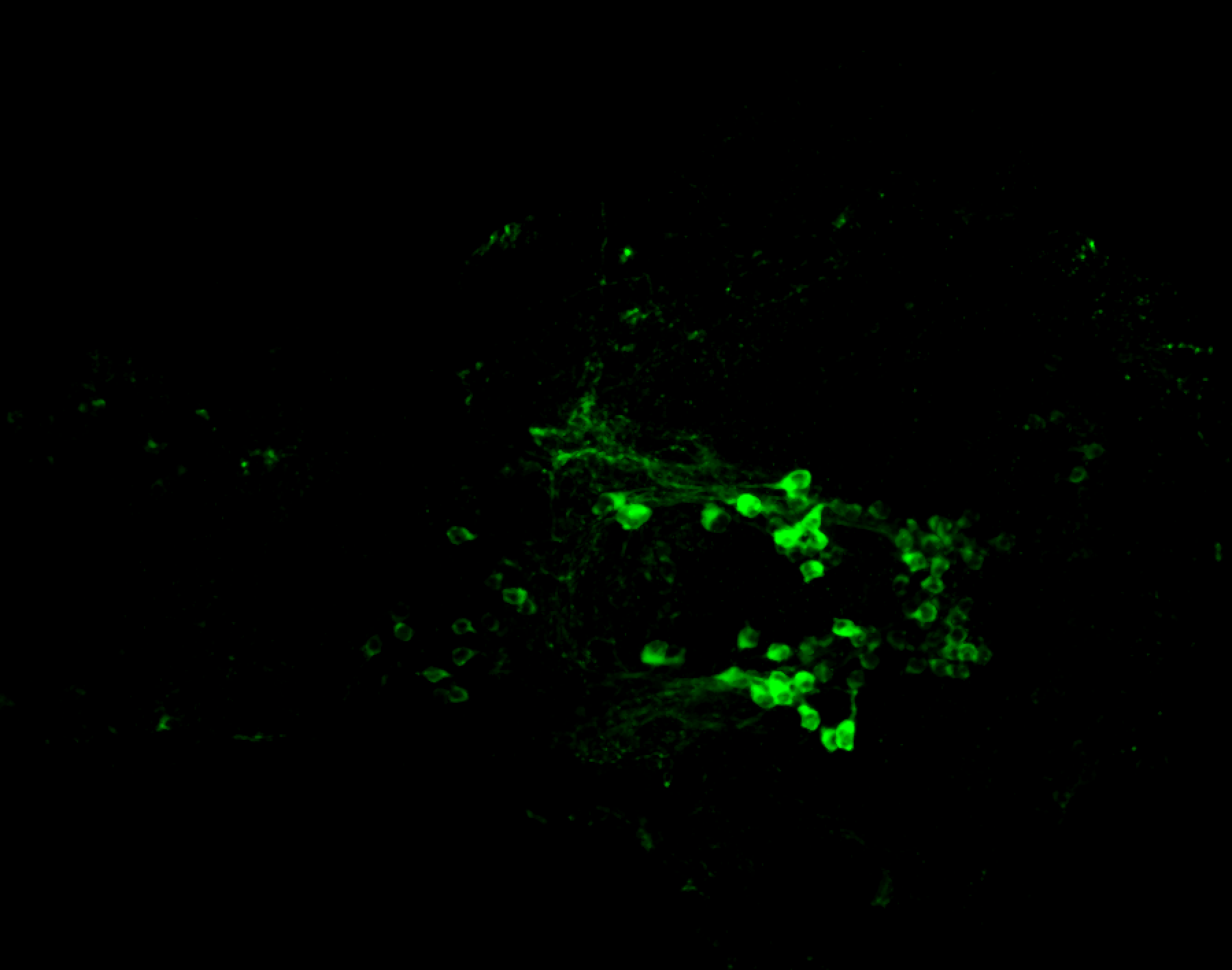
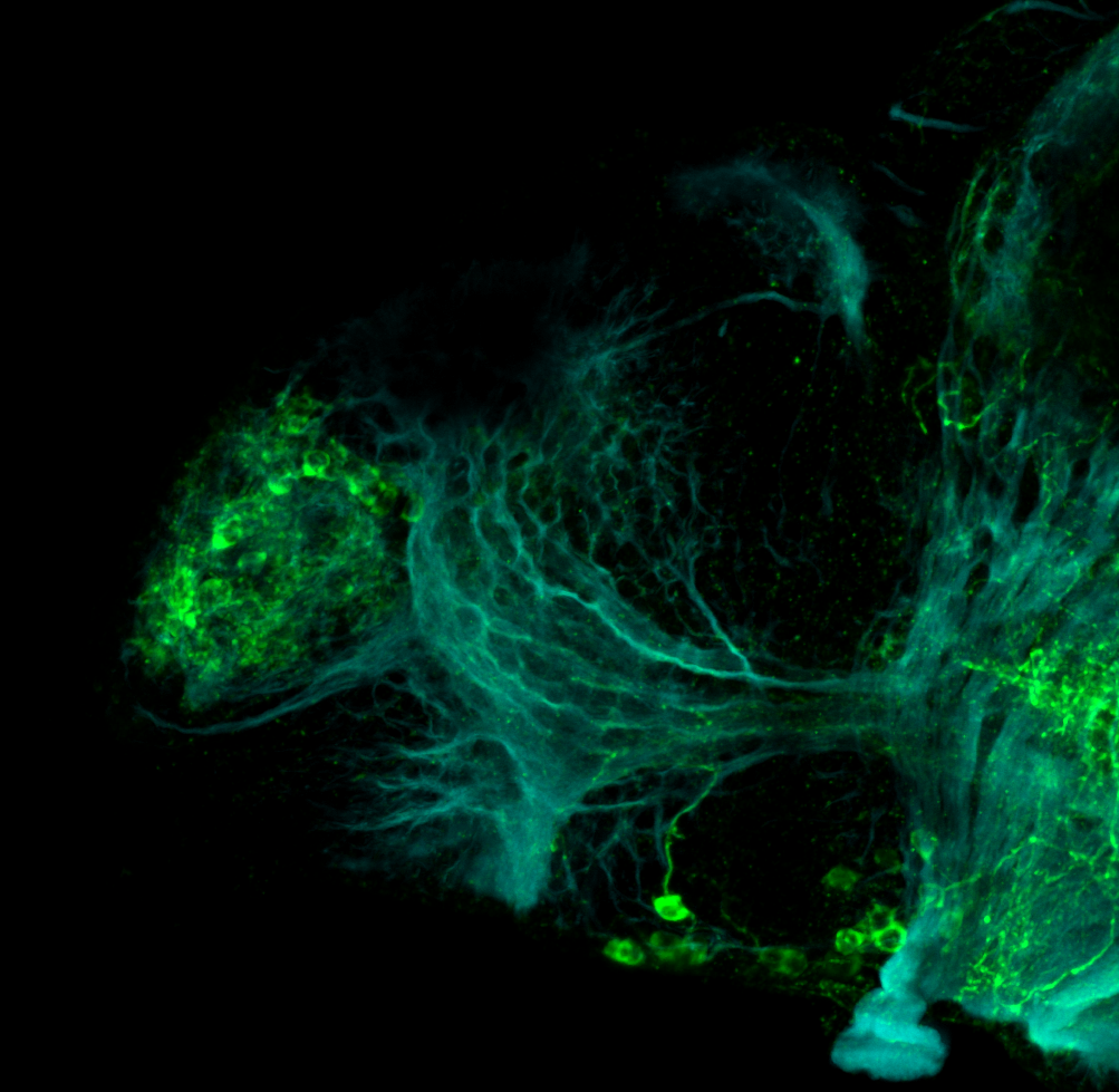
Viewing entries in
antibody

3A10

3A10

3A10

ABOUT THIS ANTIBODY

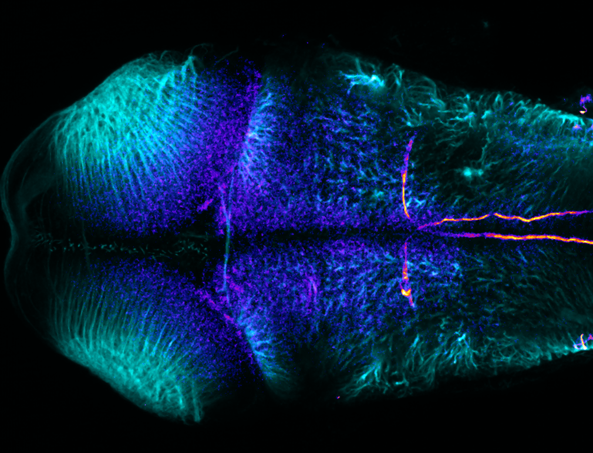
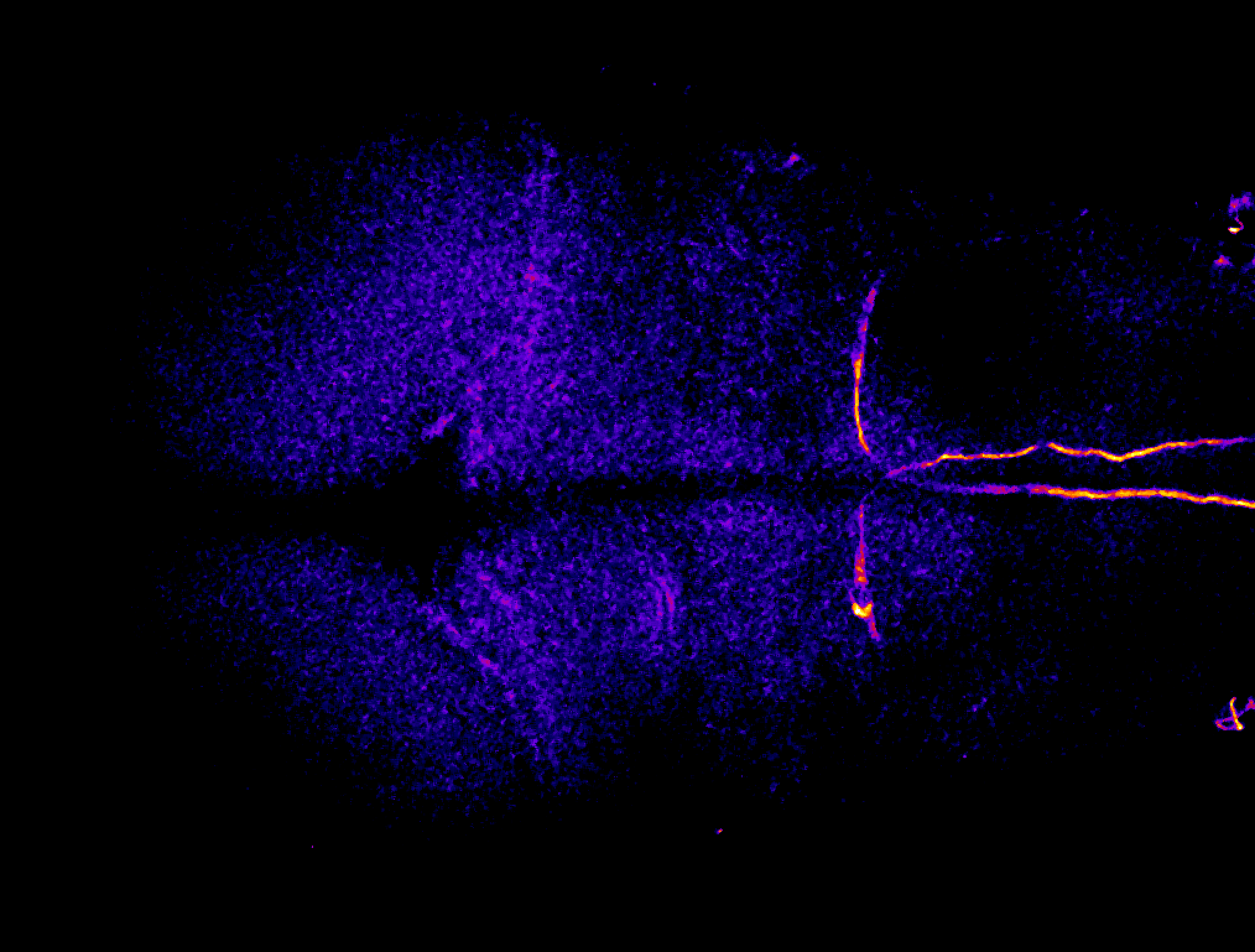
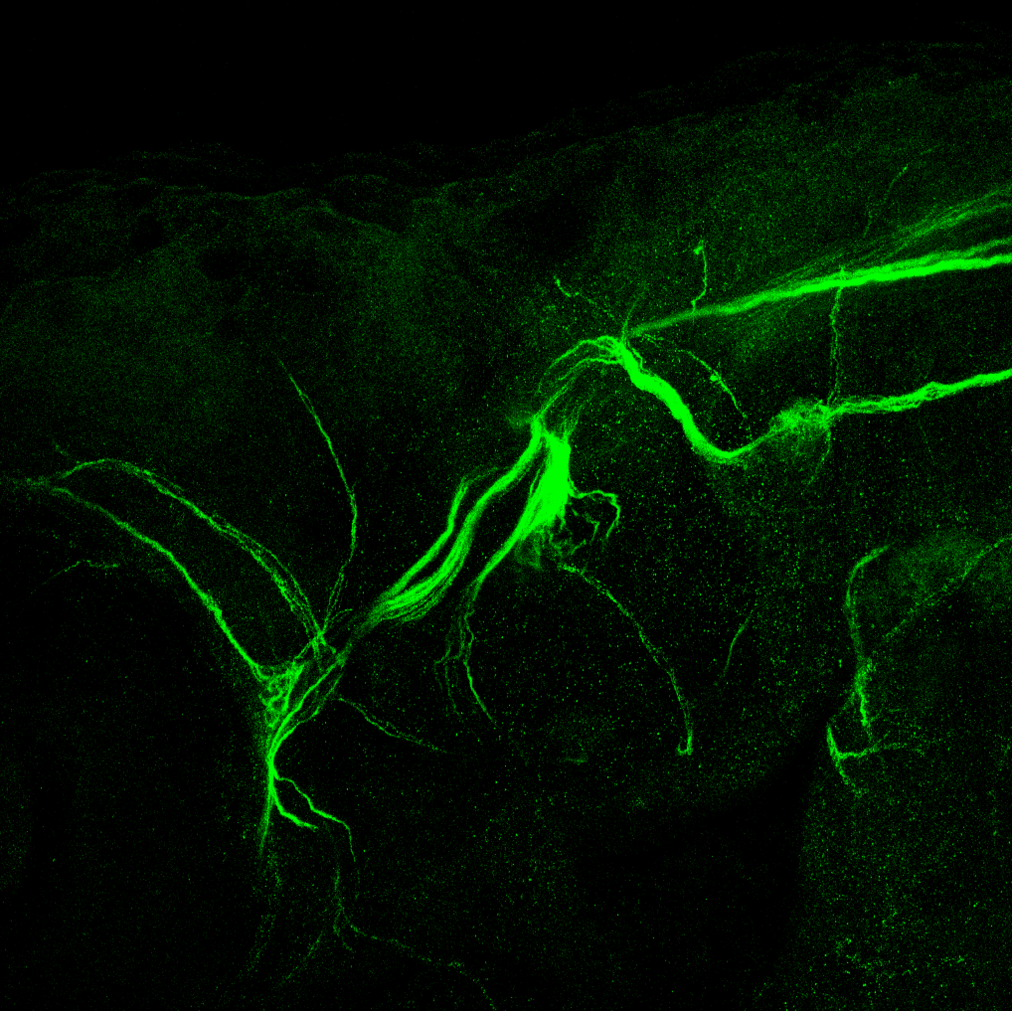
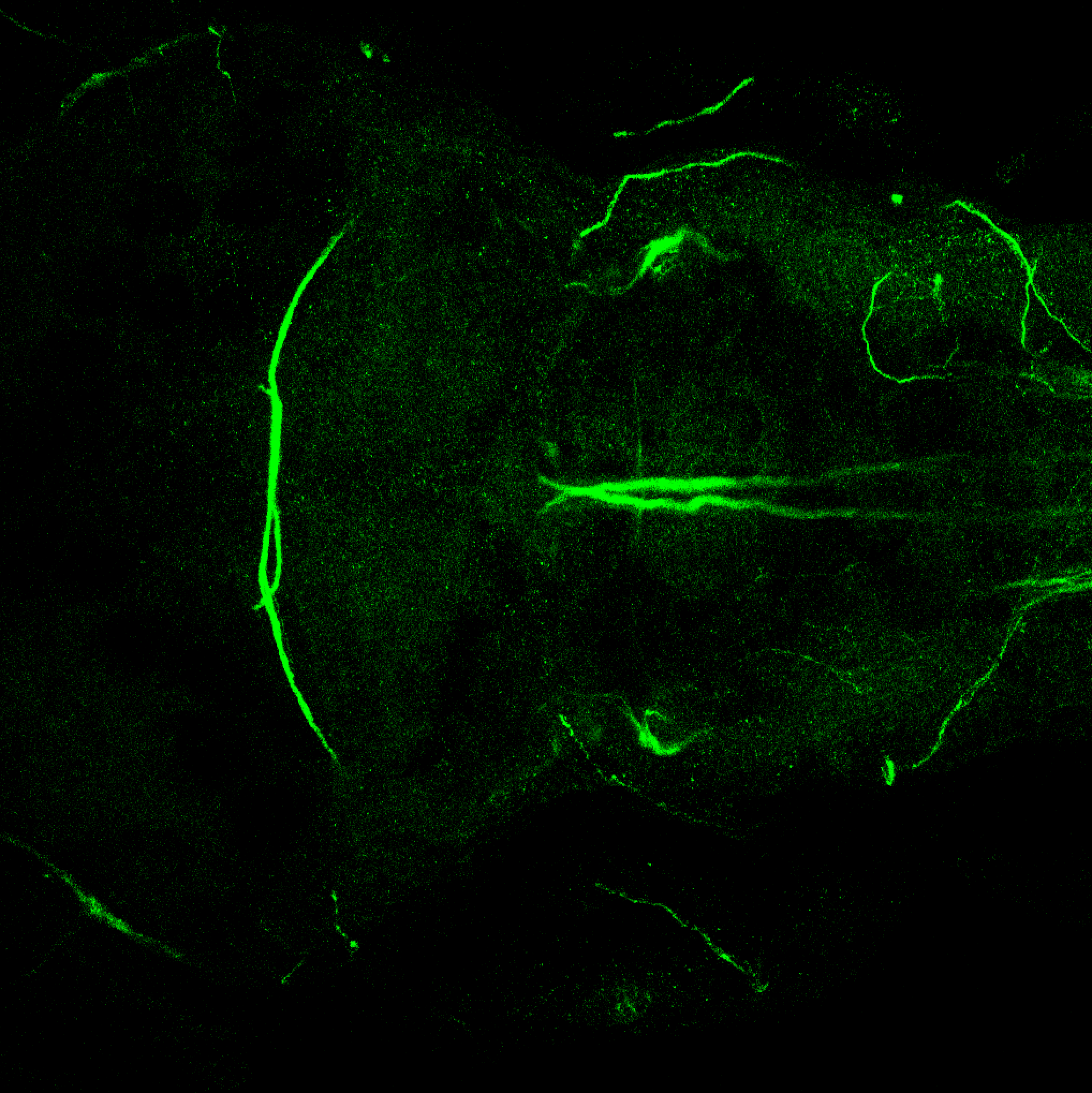
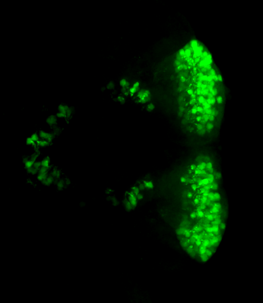
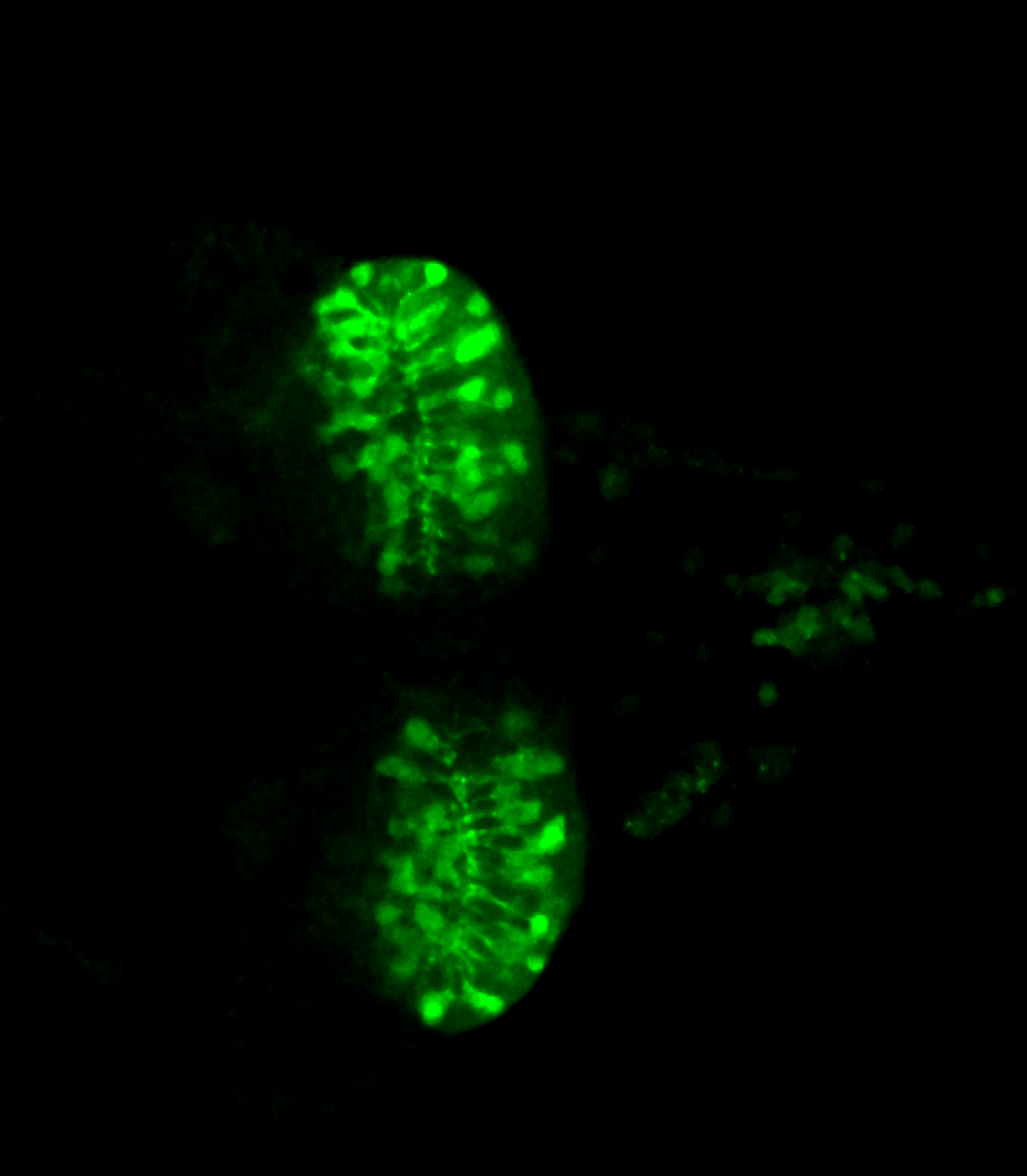
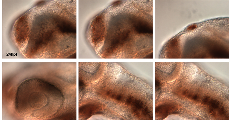
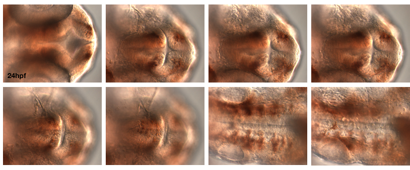
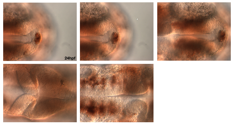
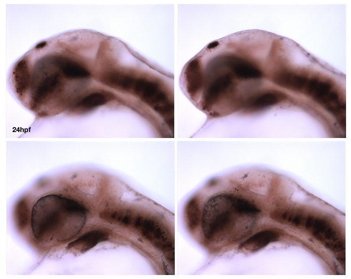
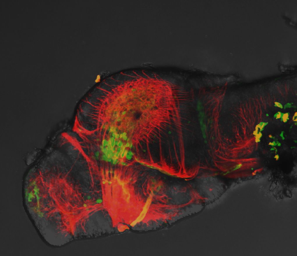
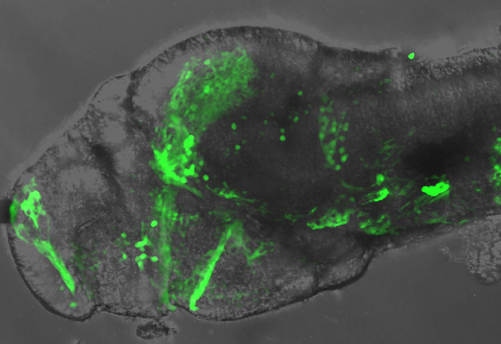
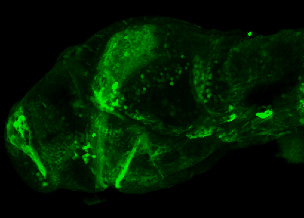
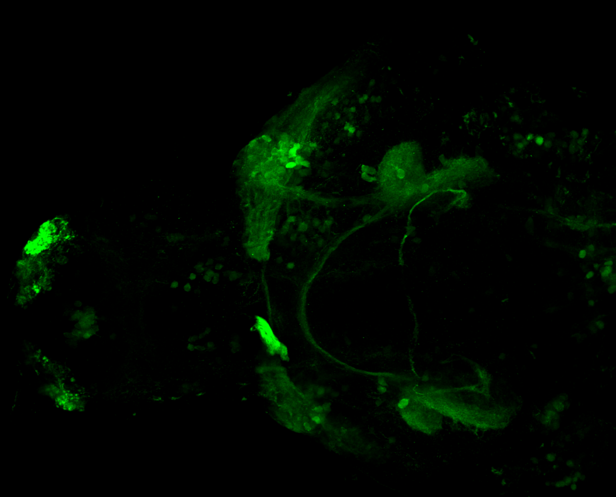
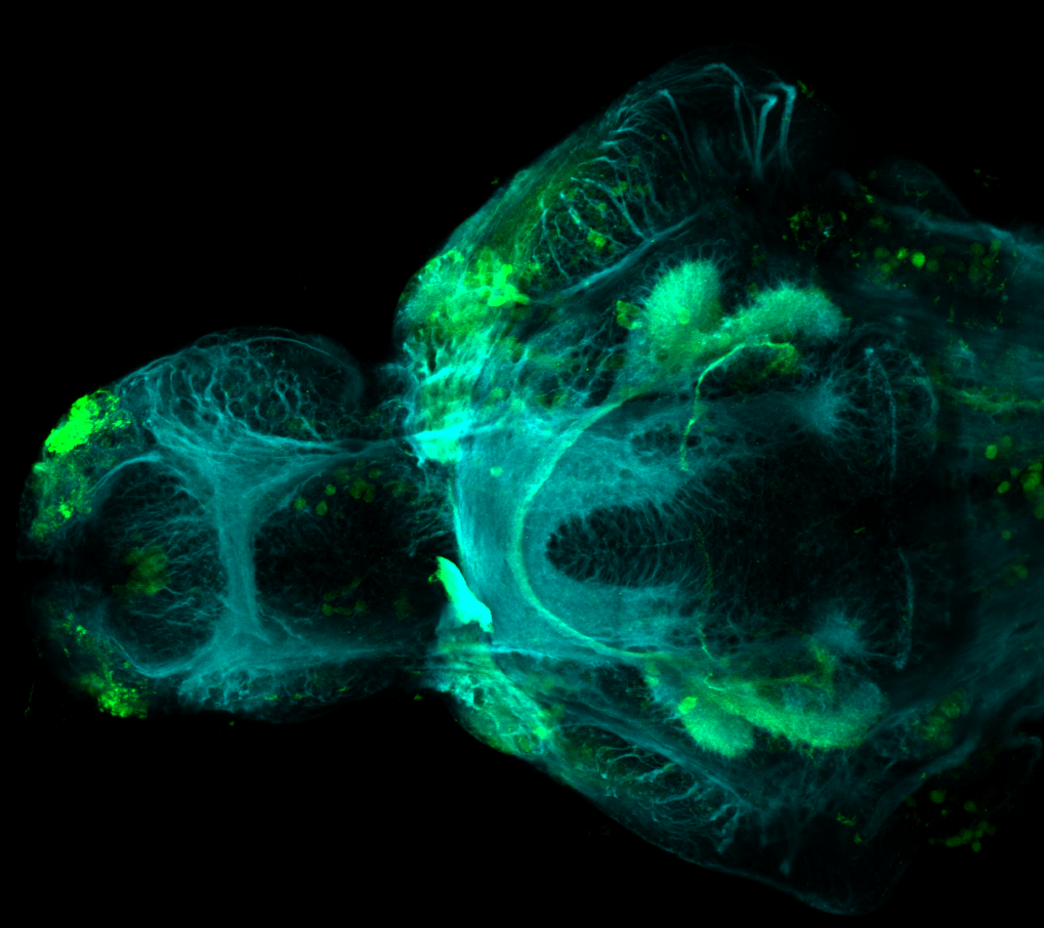
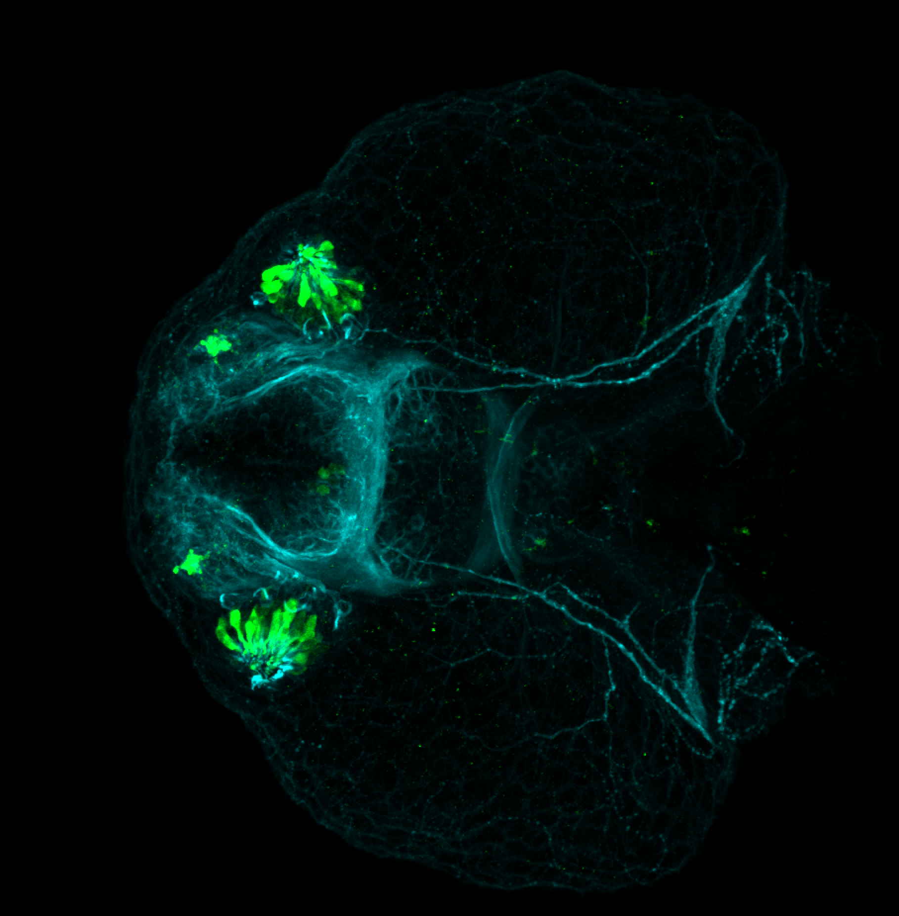
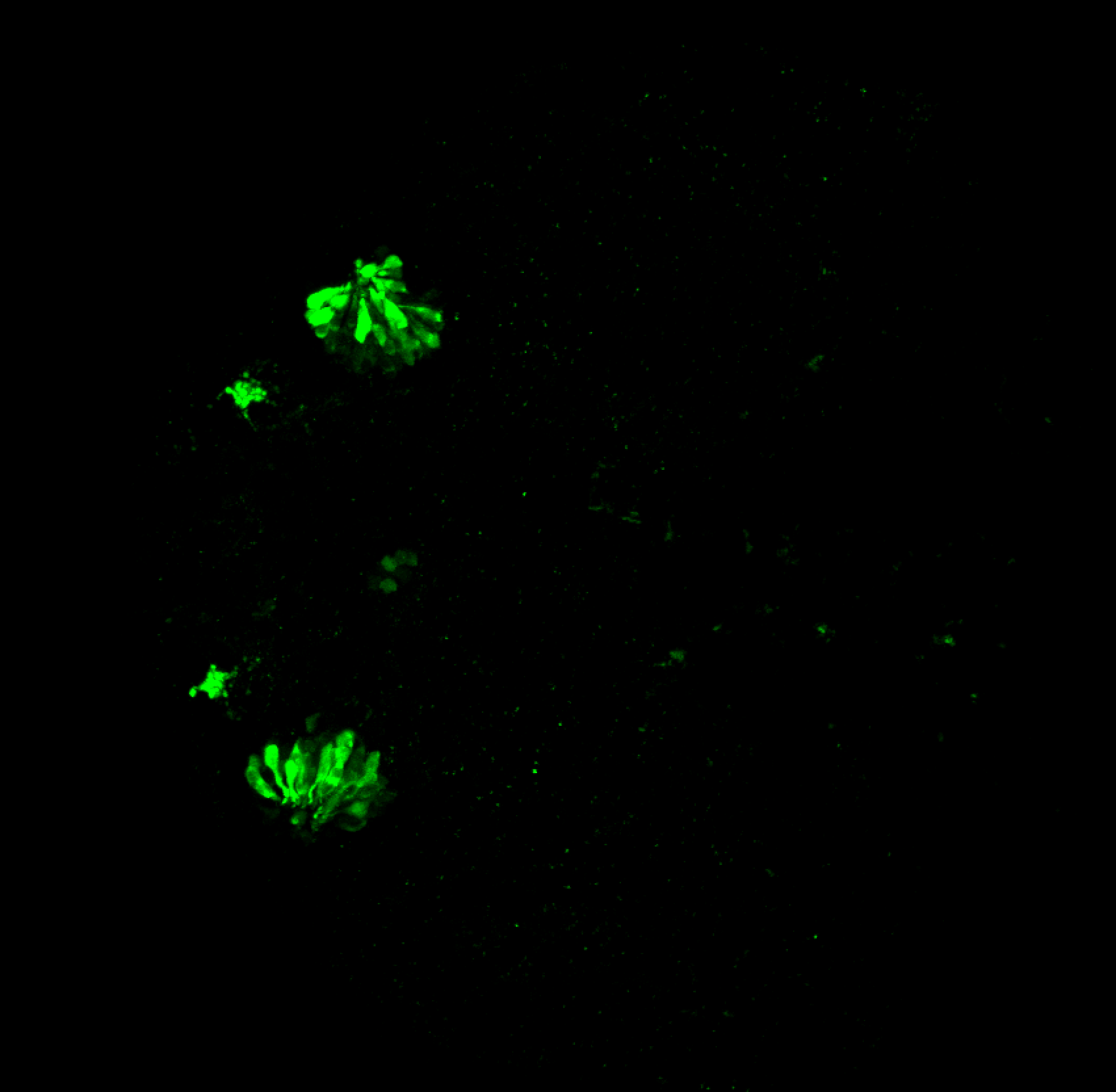
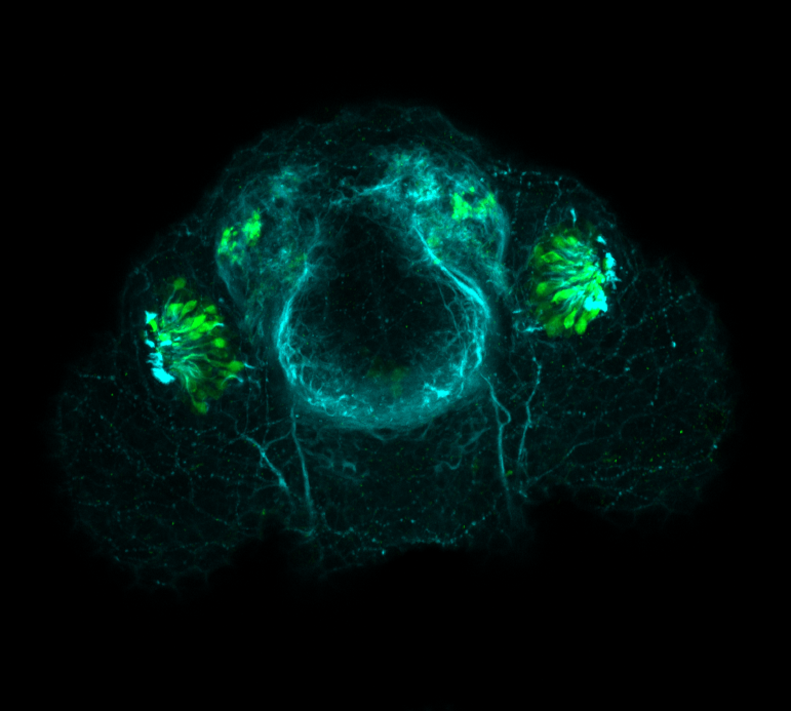
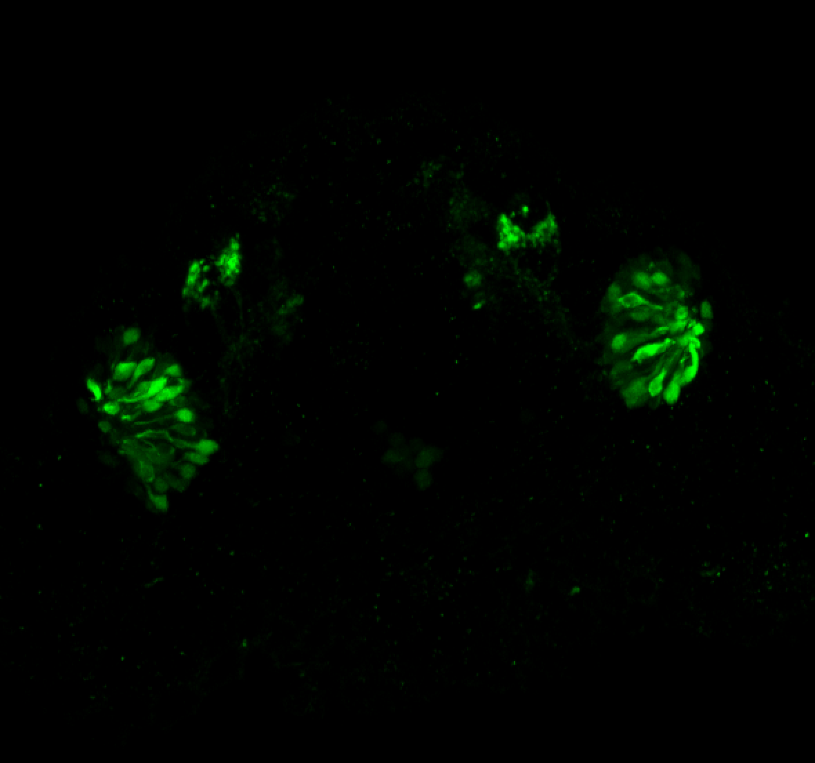
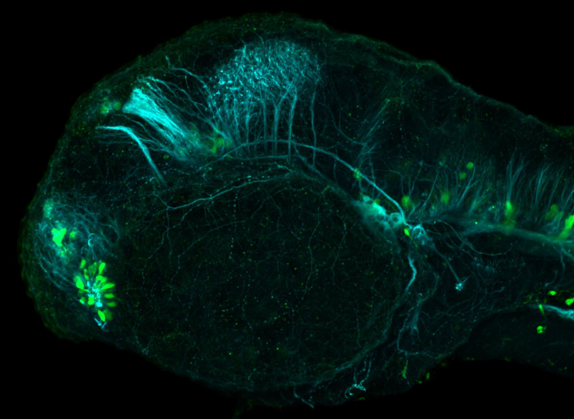
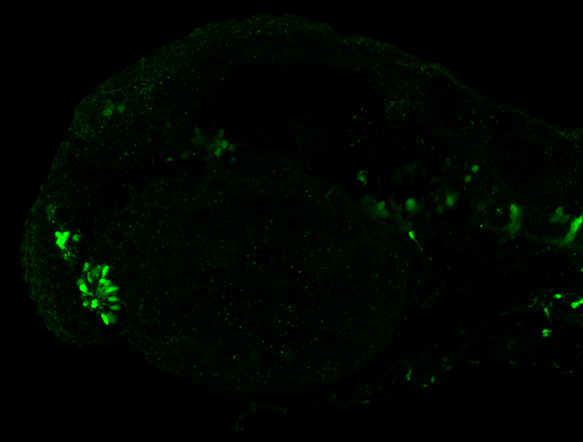
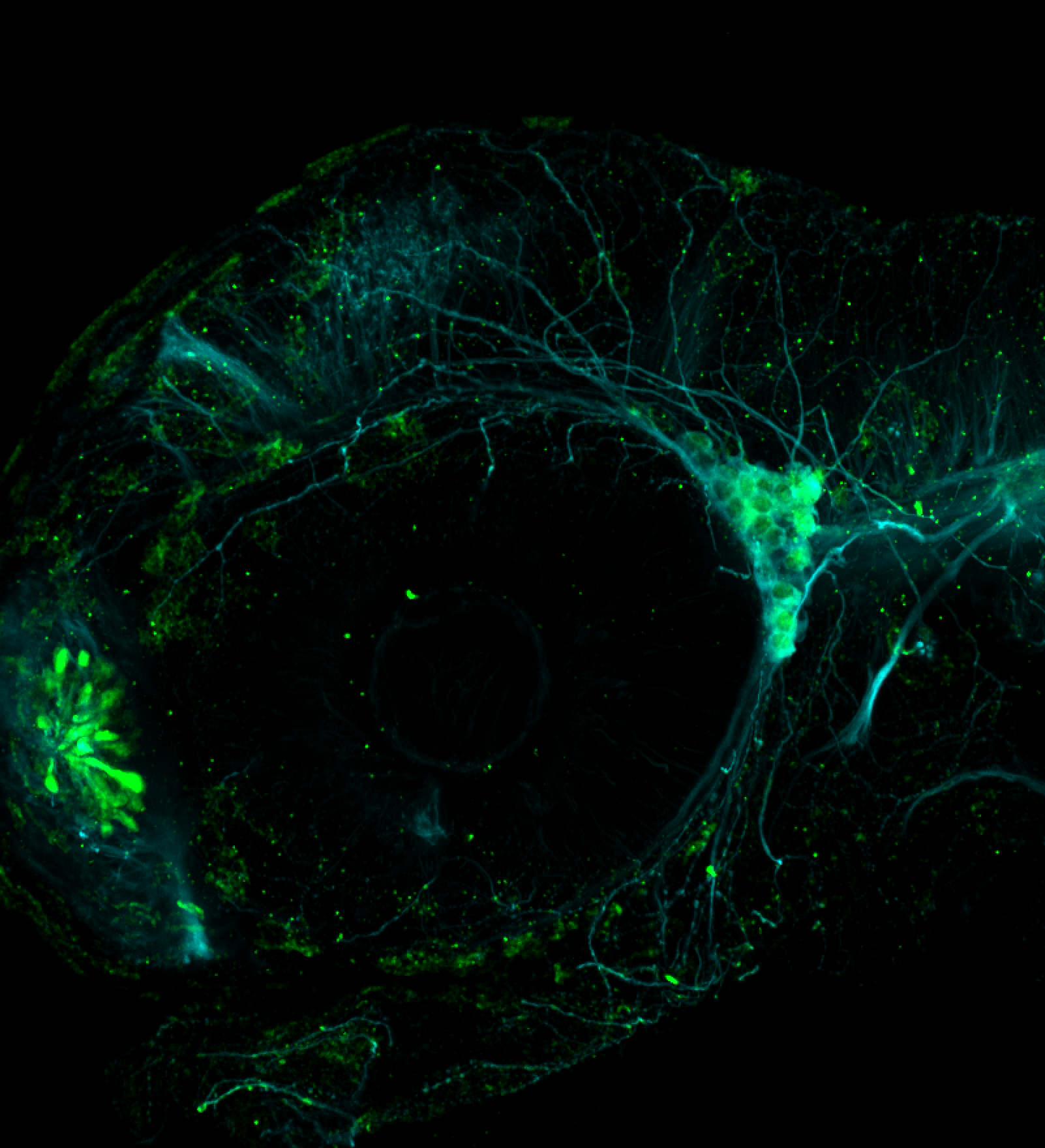
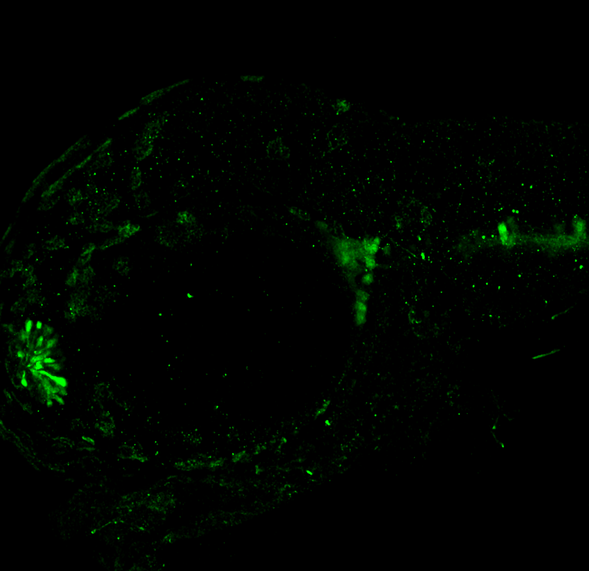
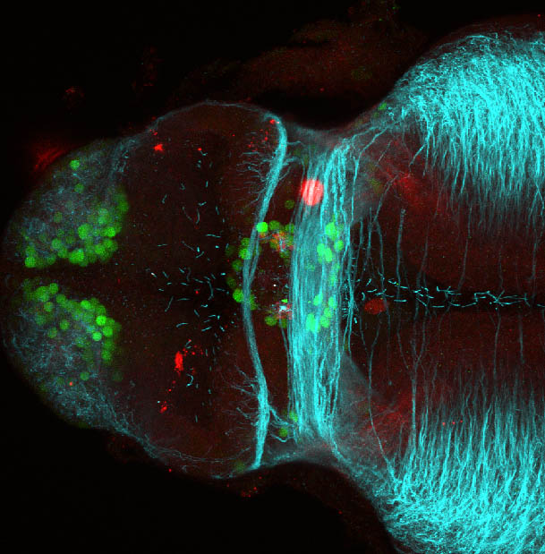
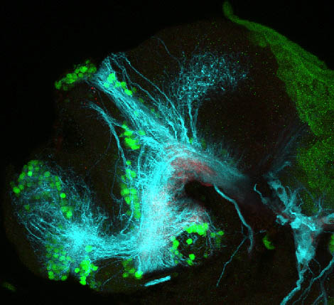
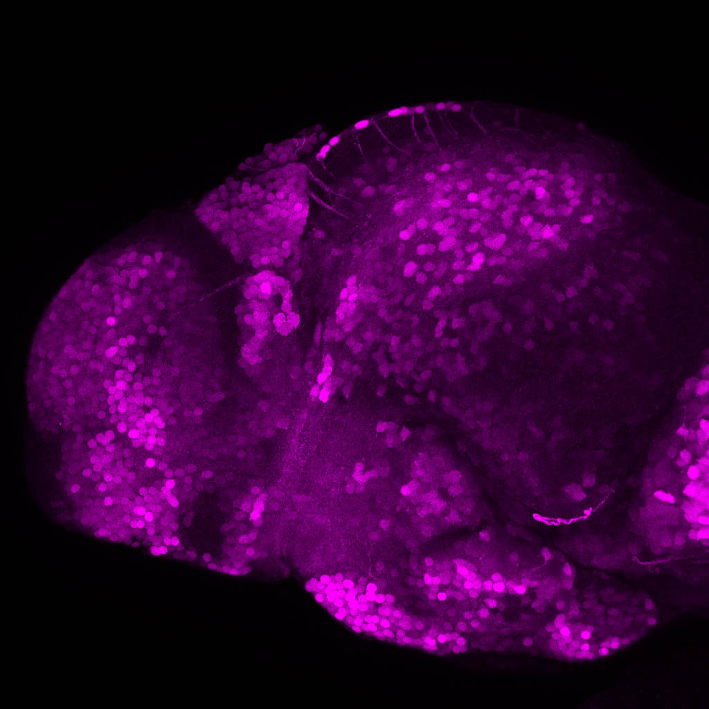
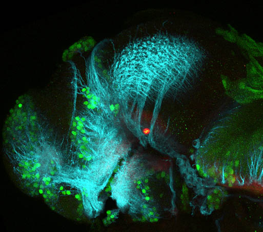
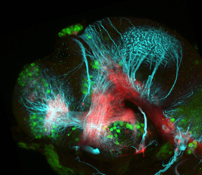
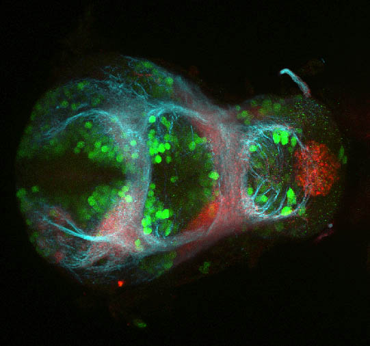
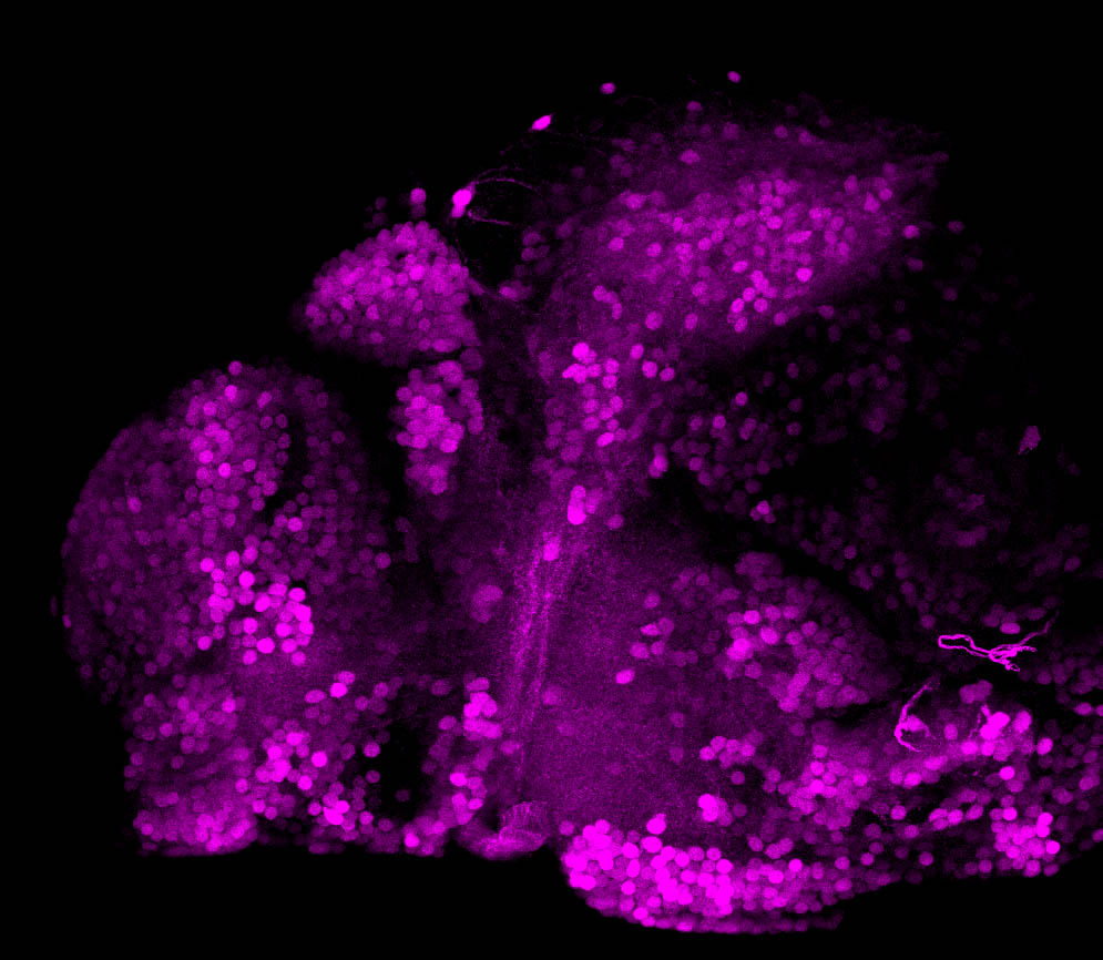
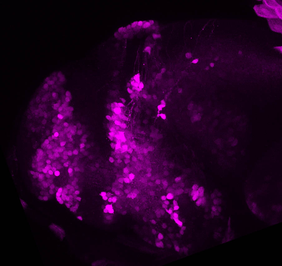
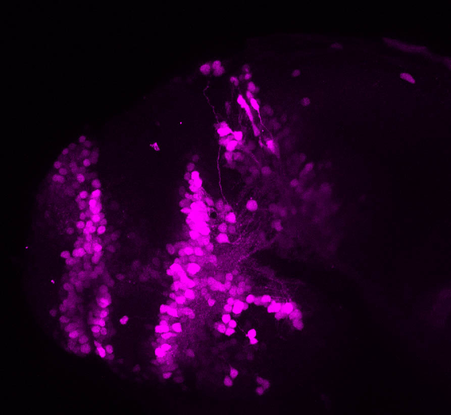
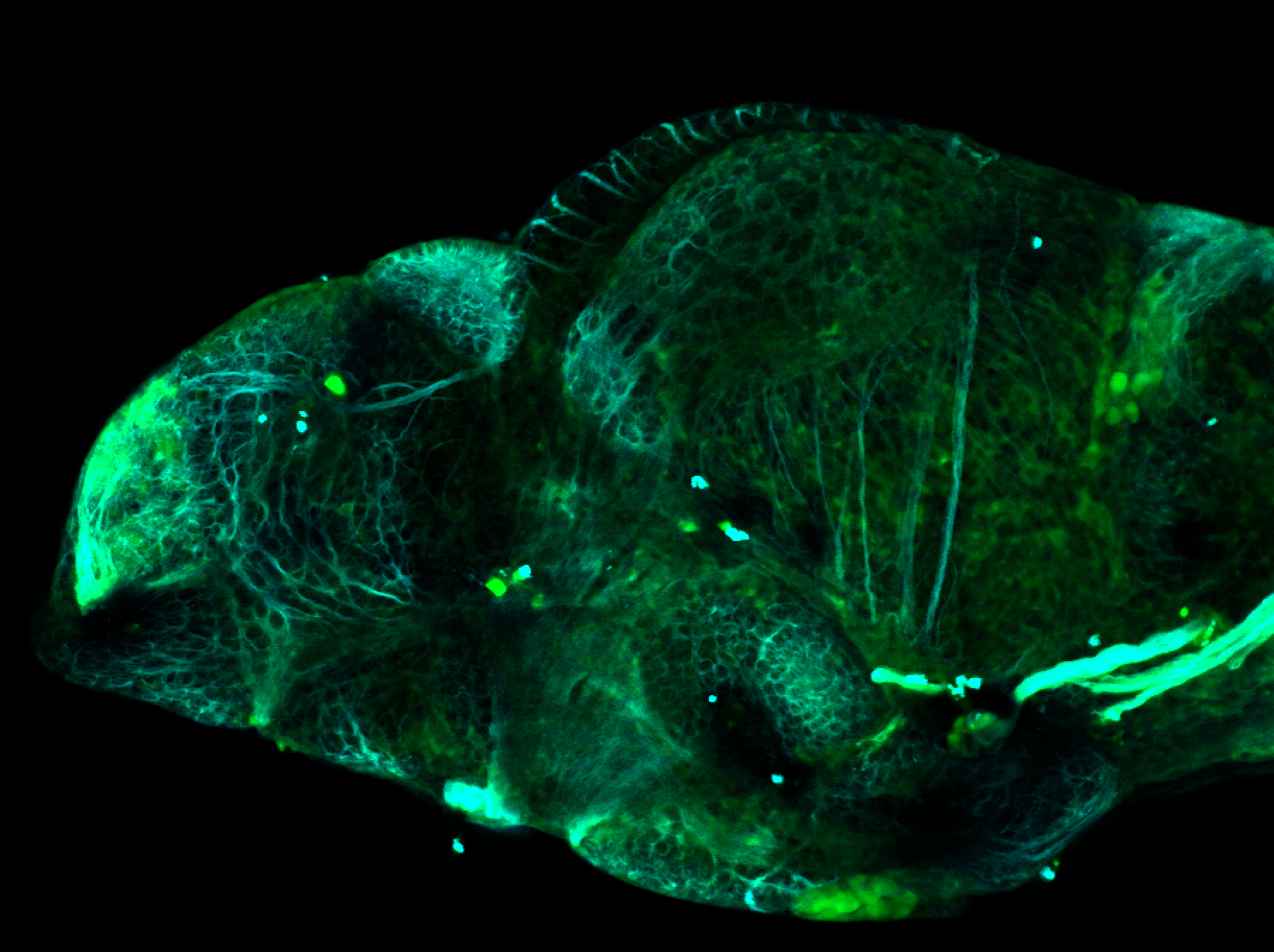
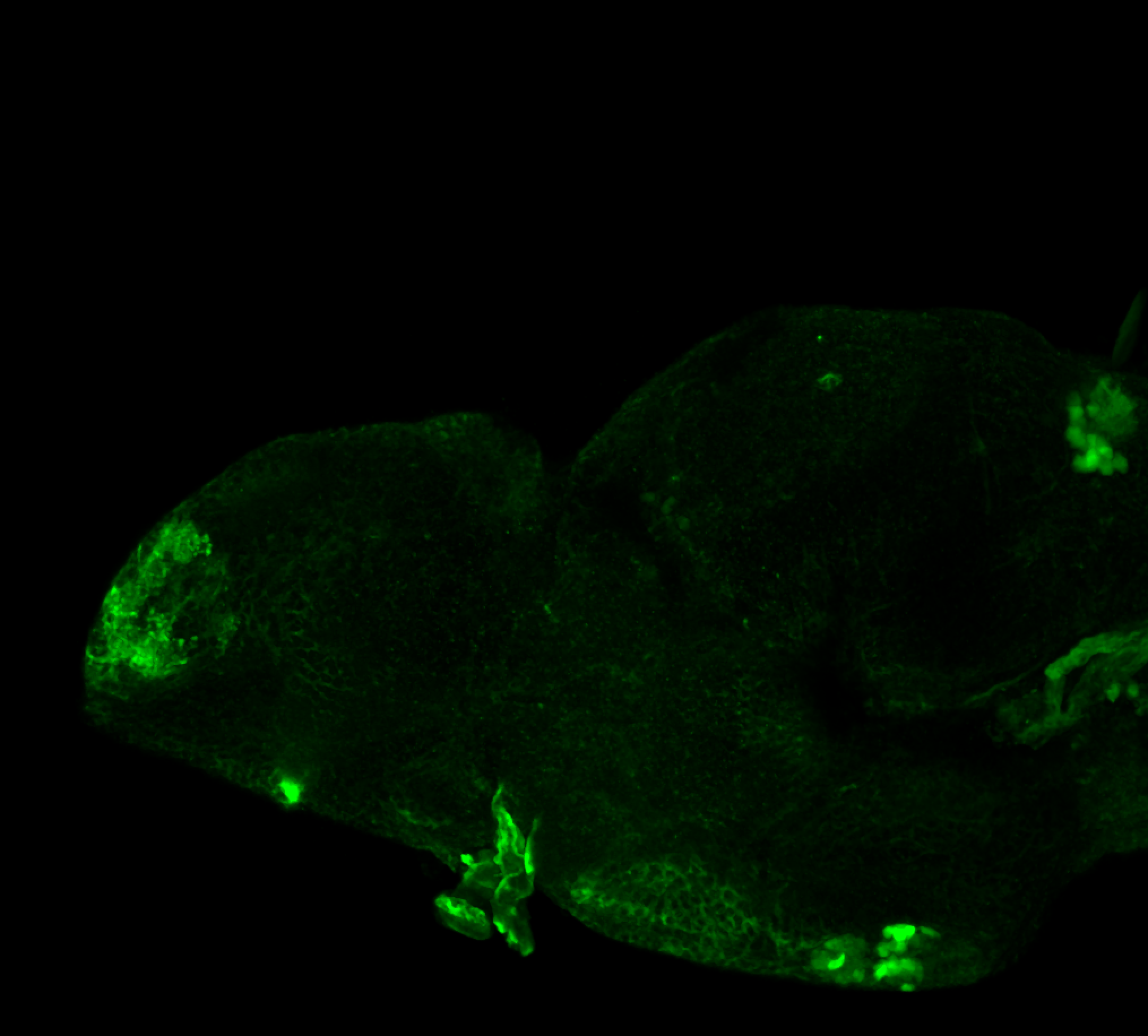
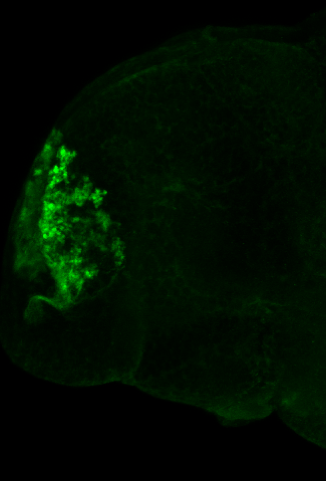
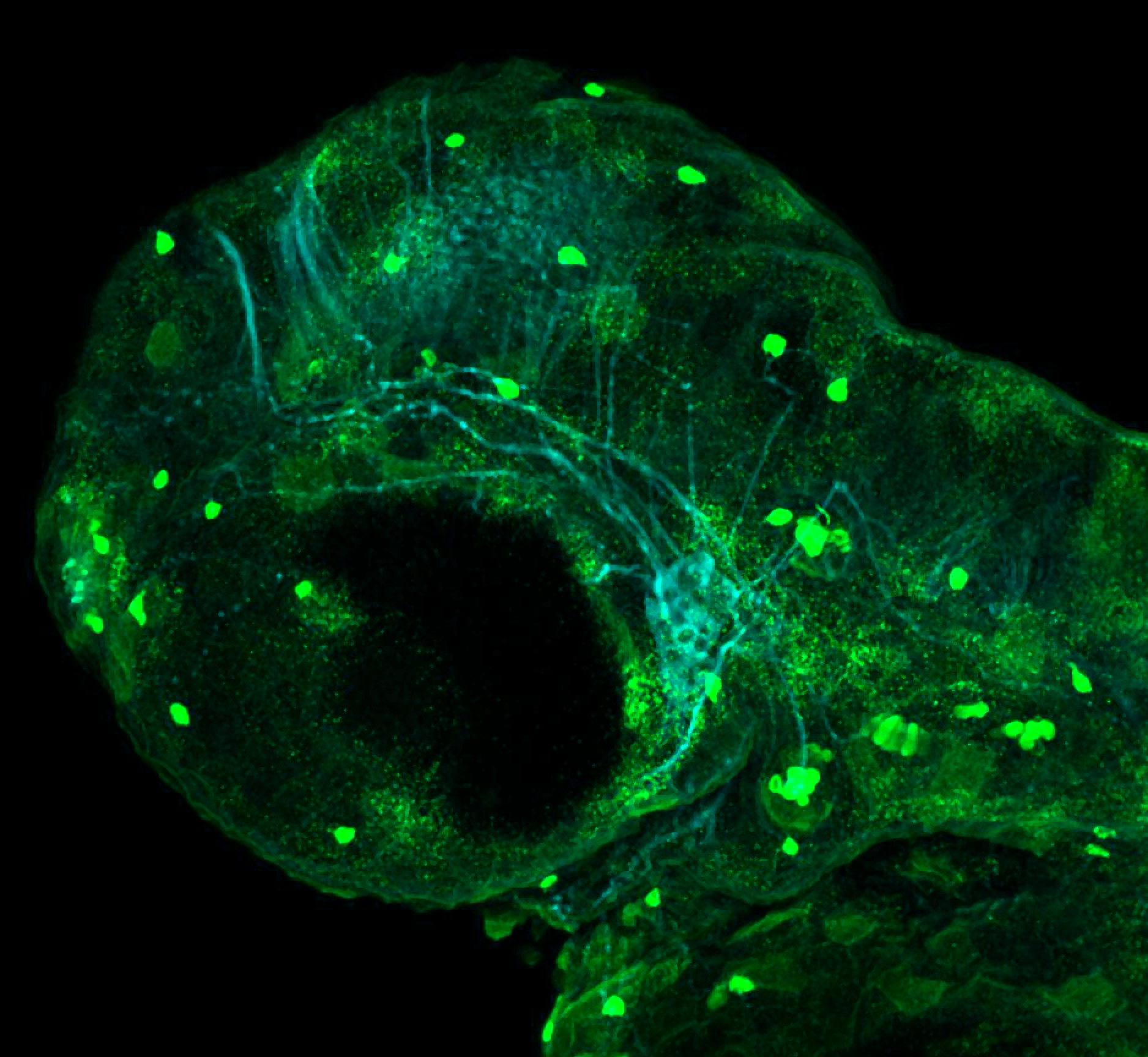
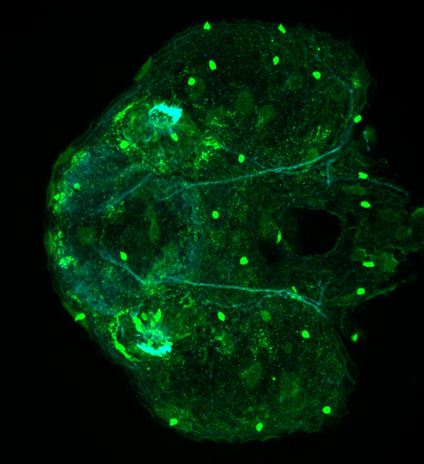
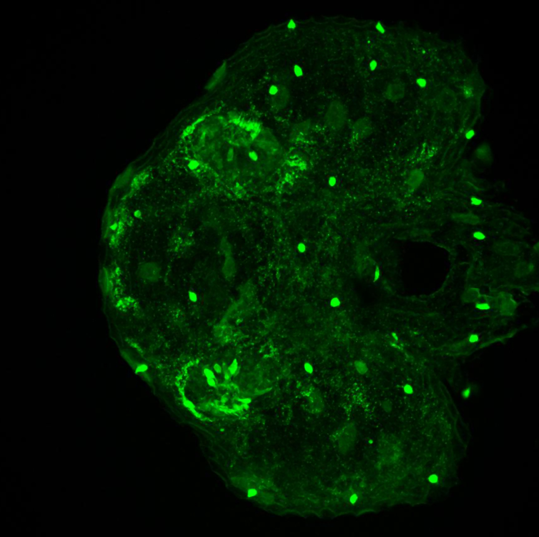
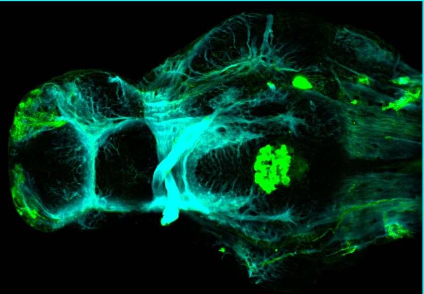
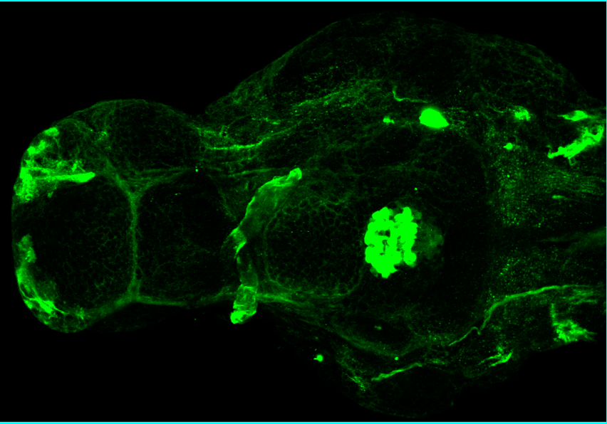
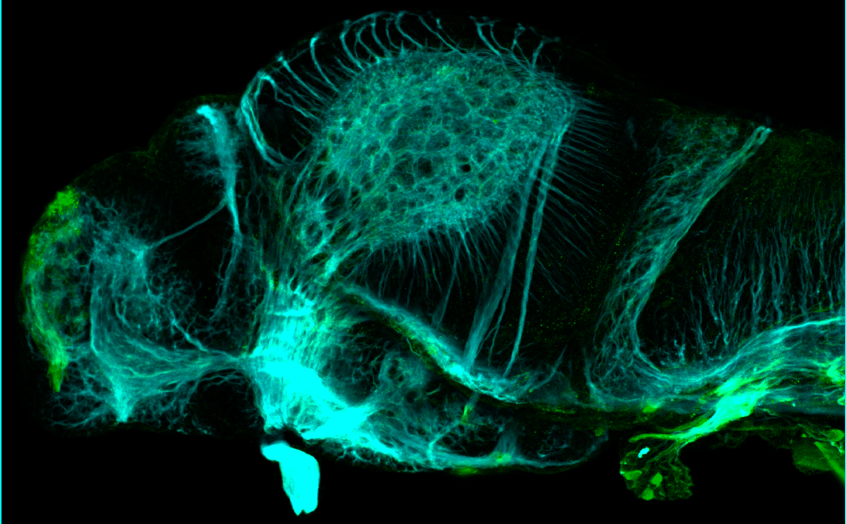
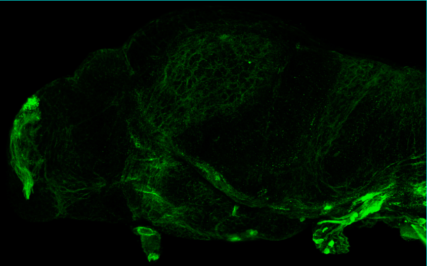
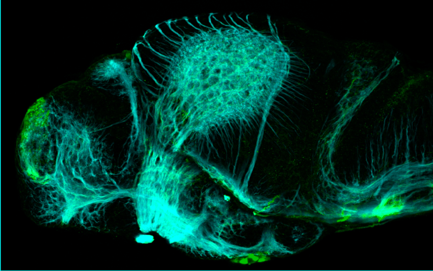
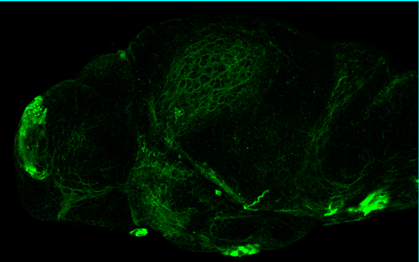
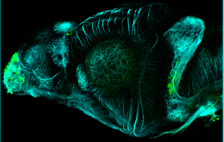
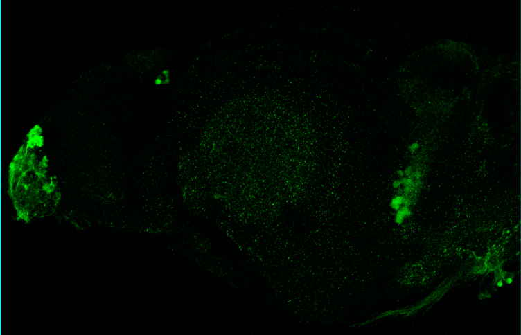
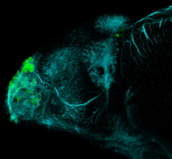
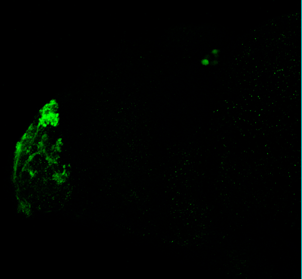
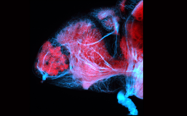
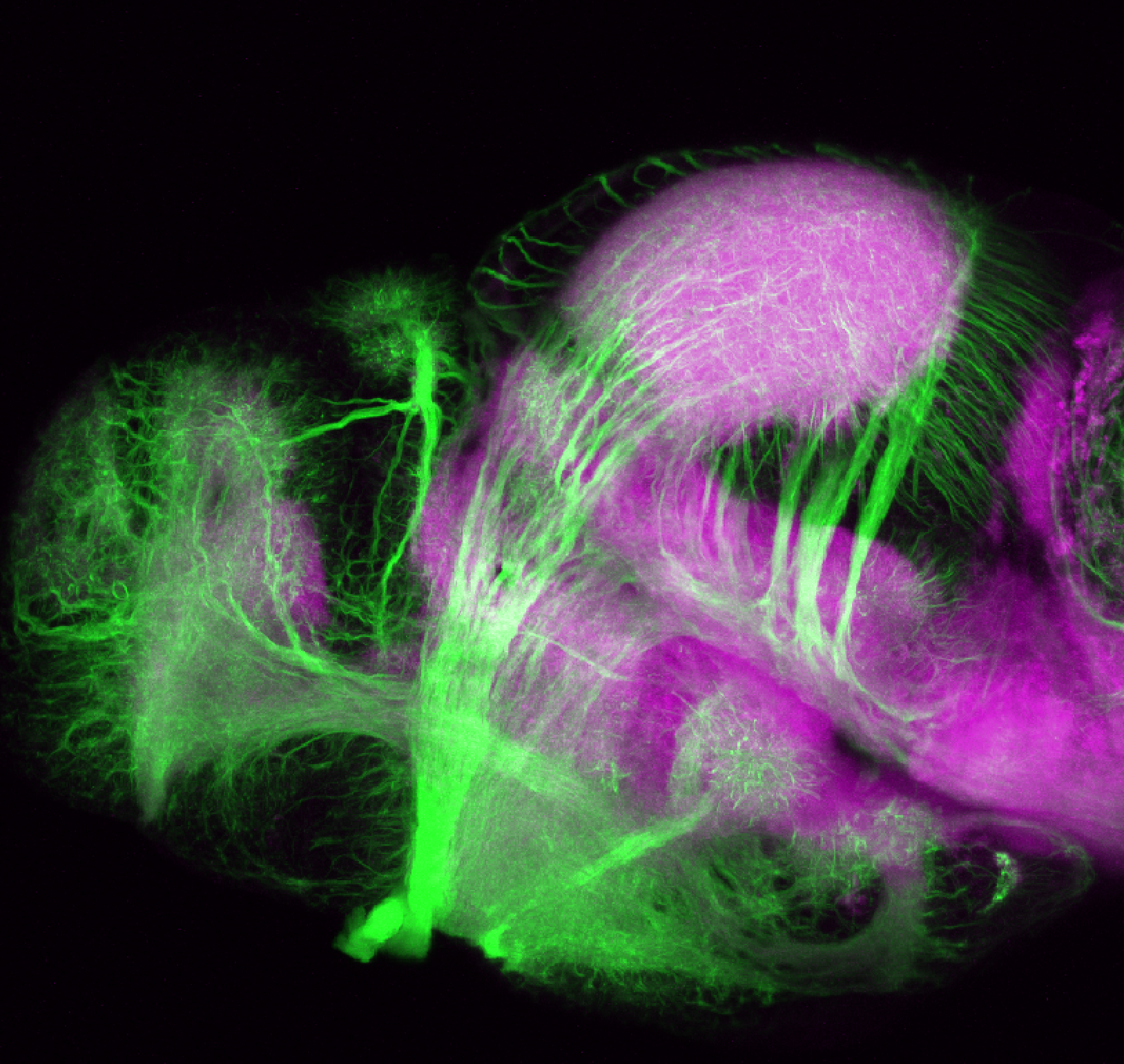
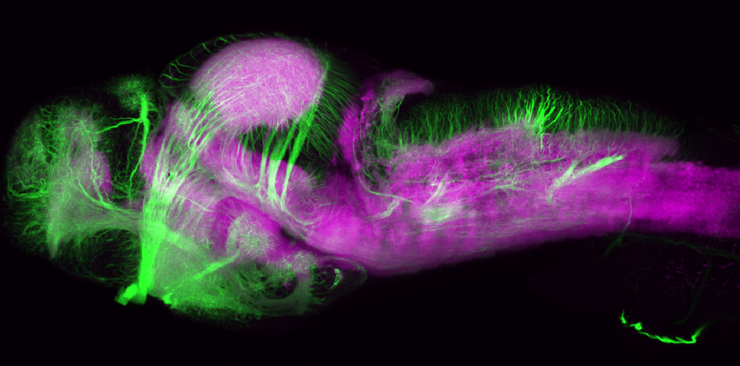
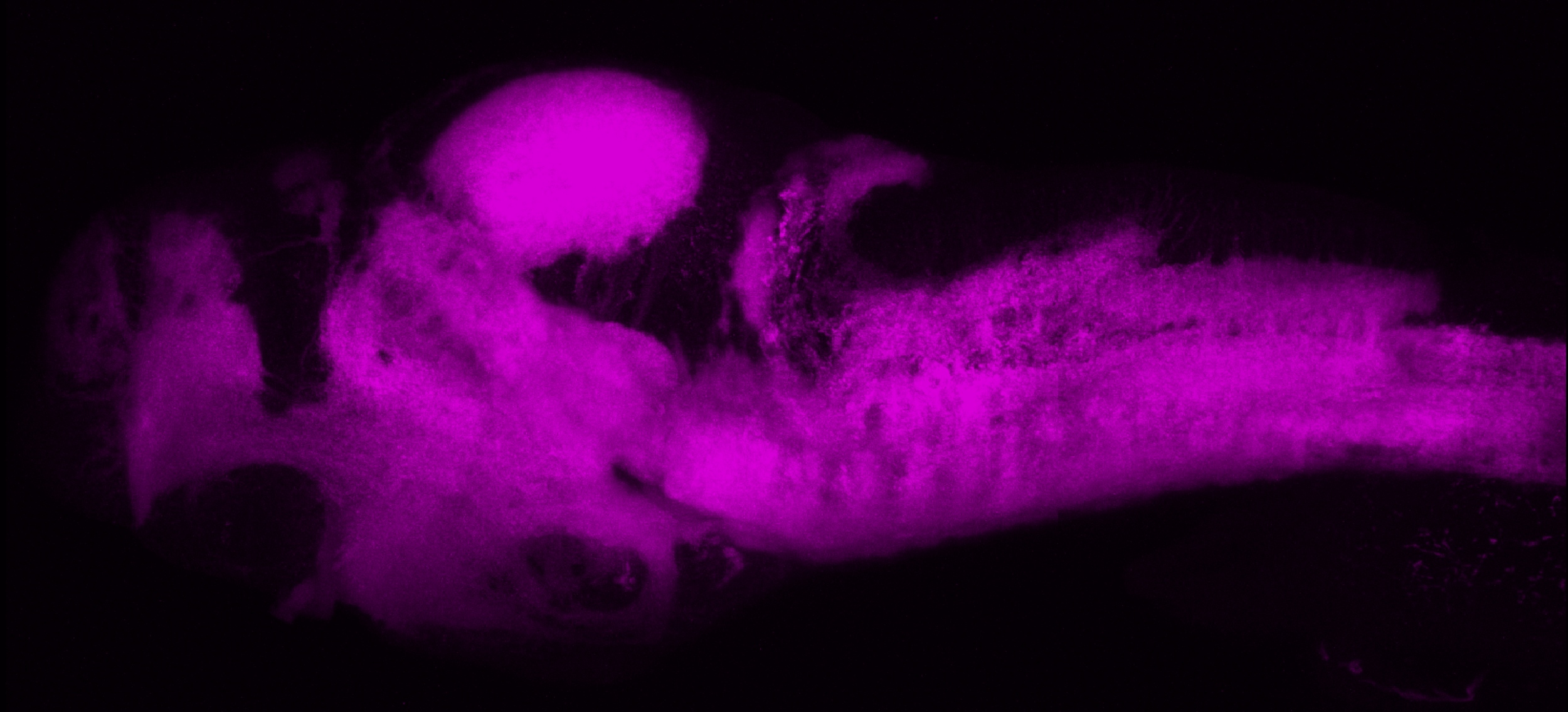
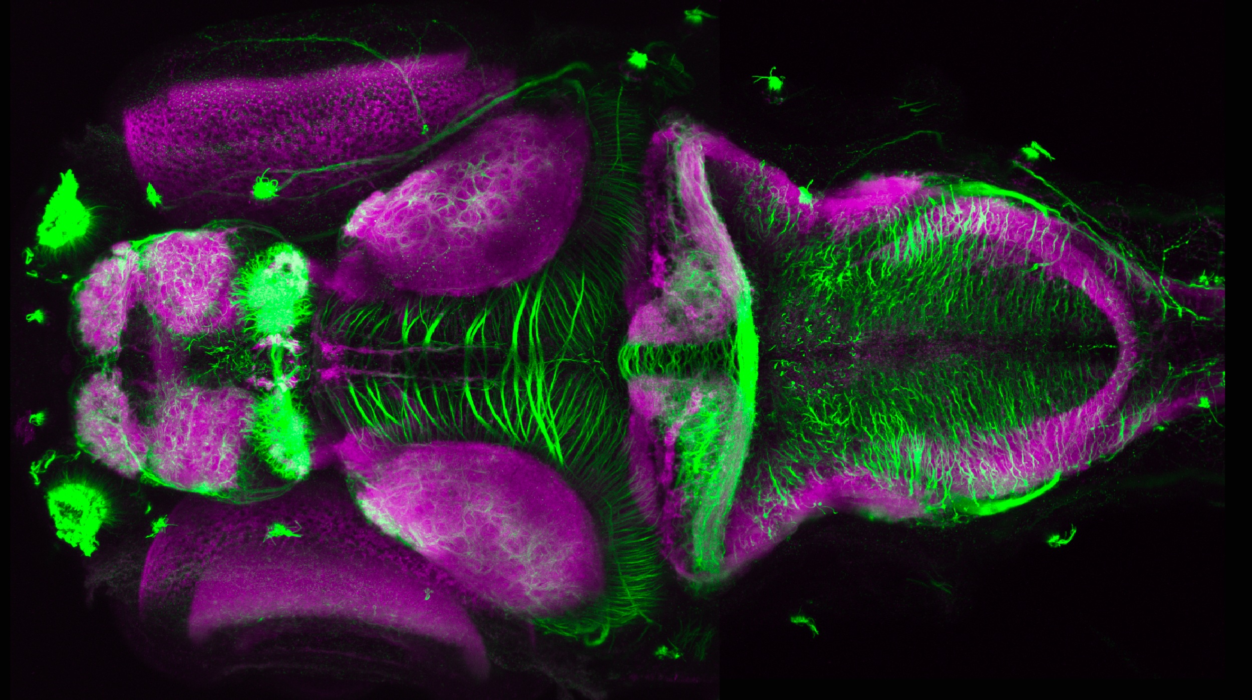
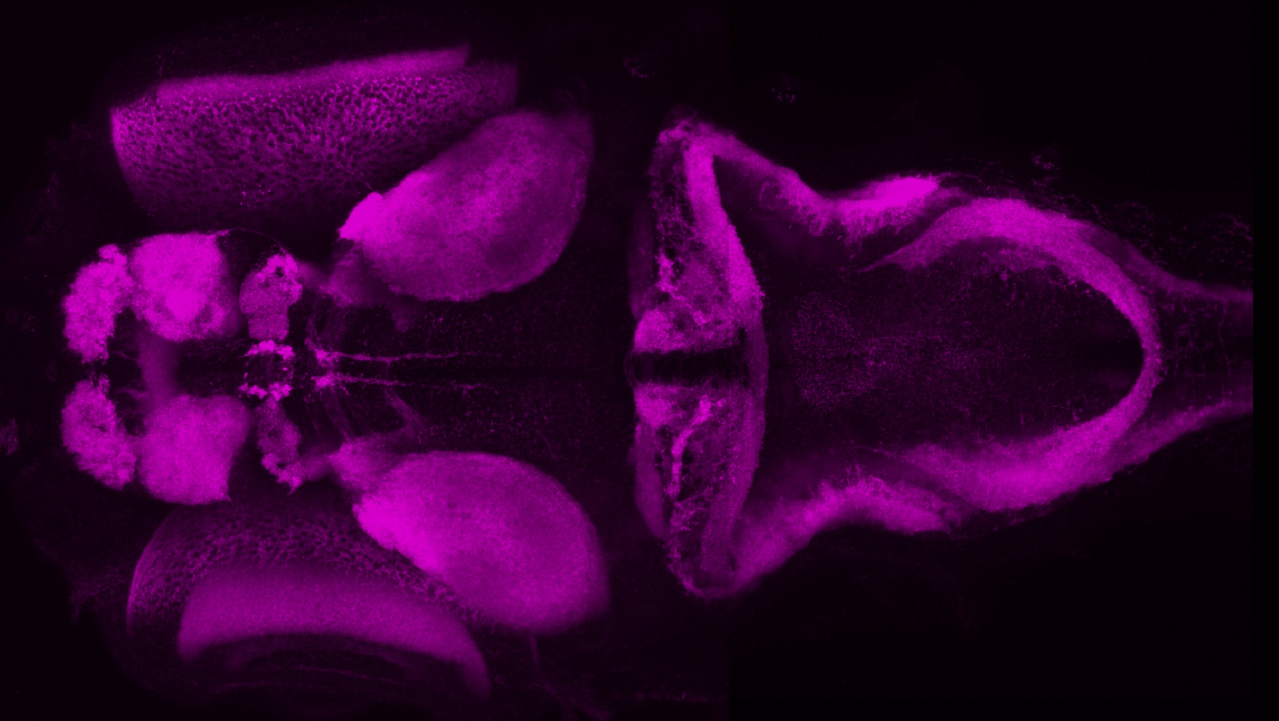
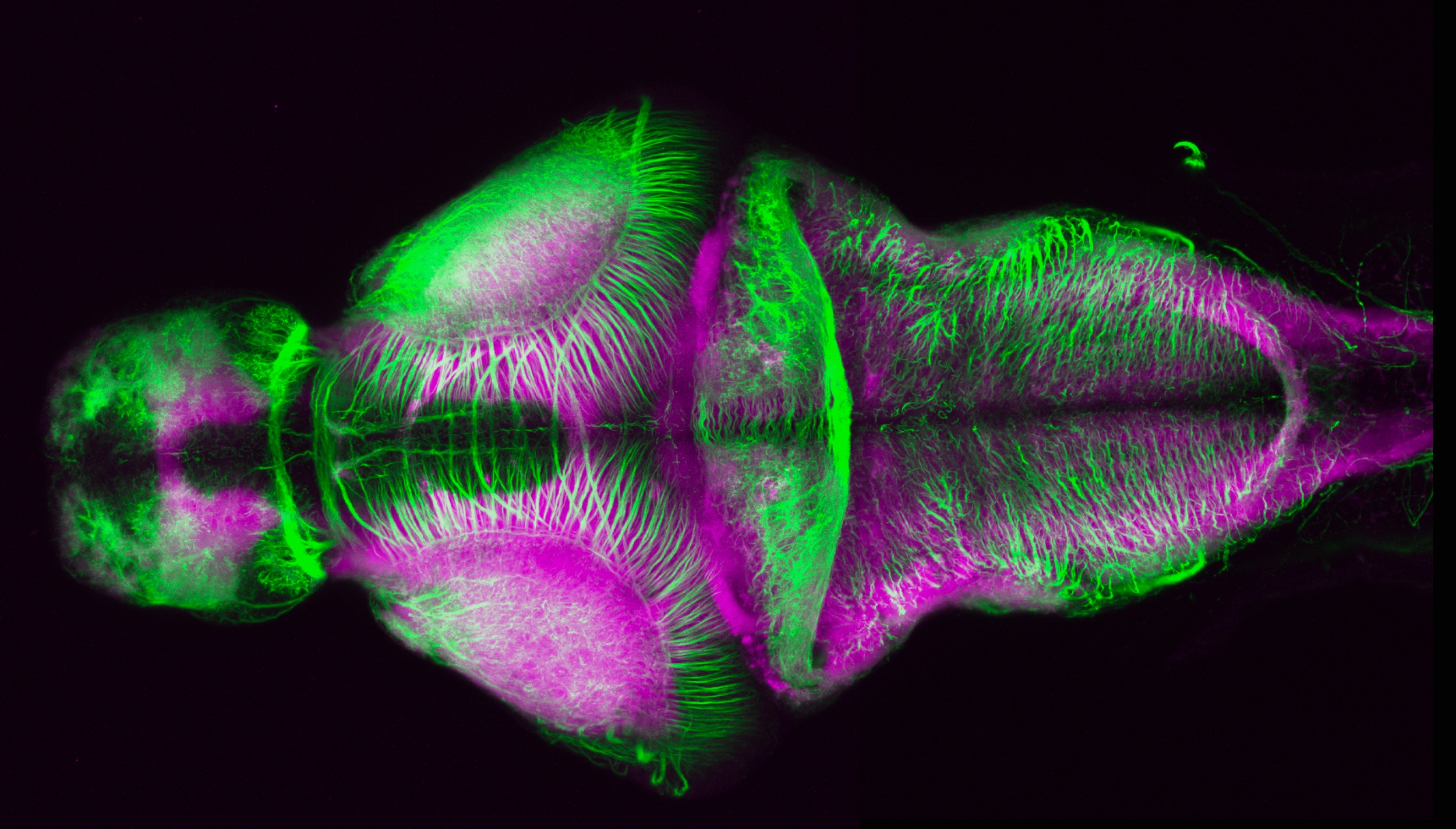
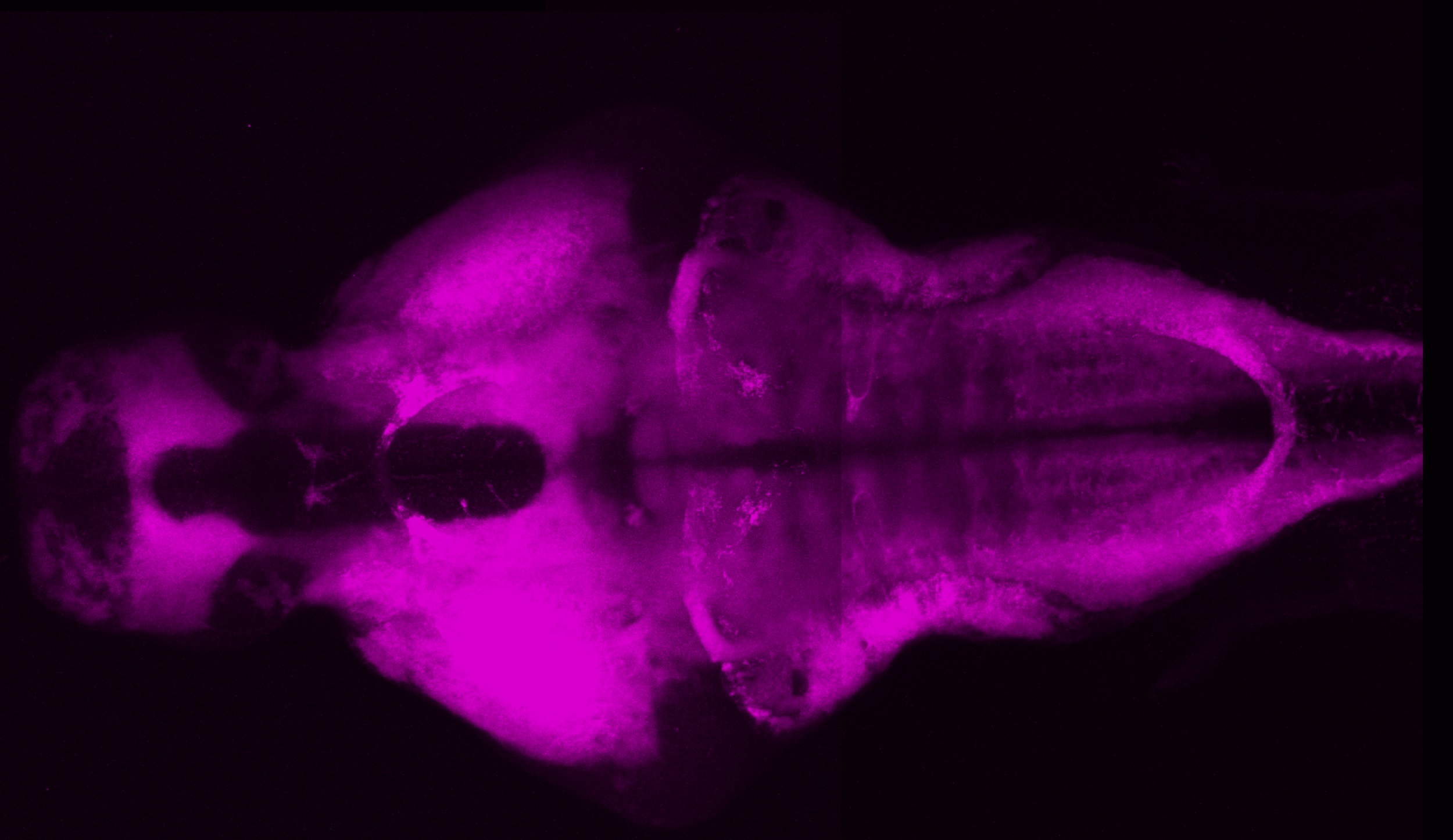
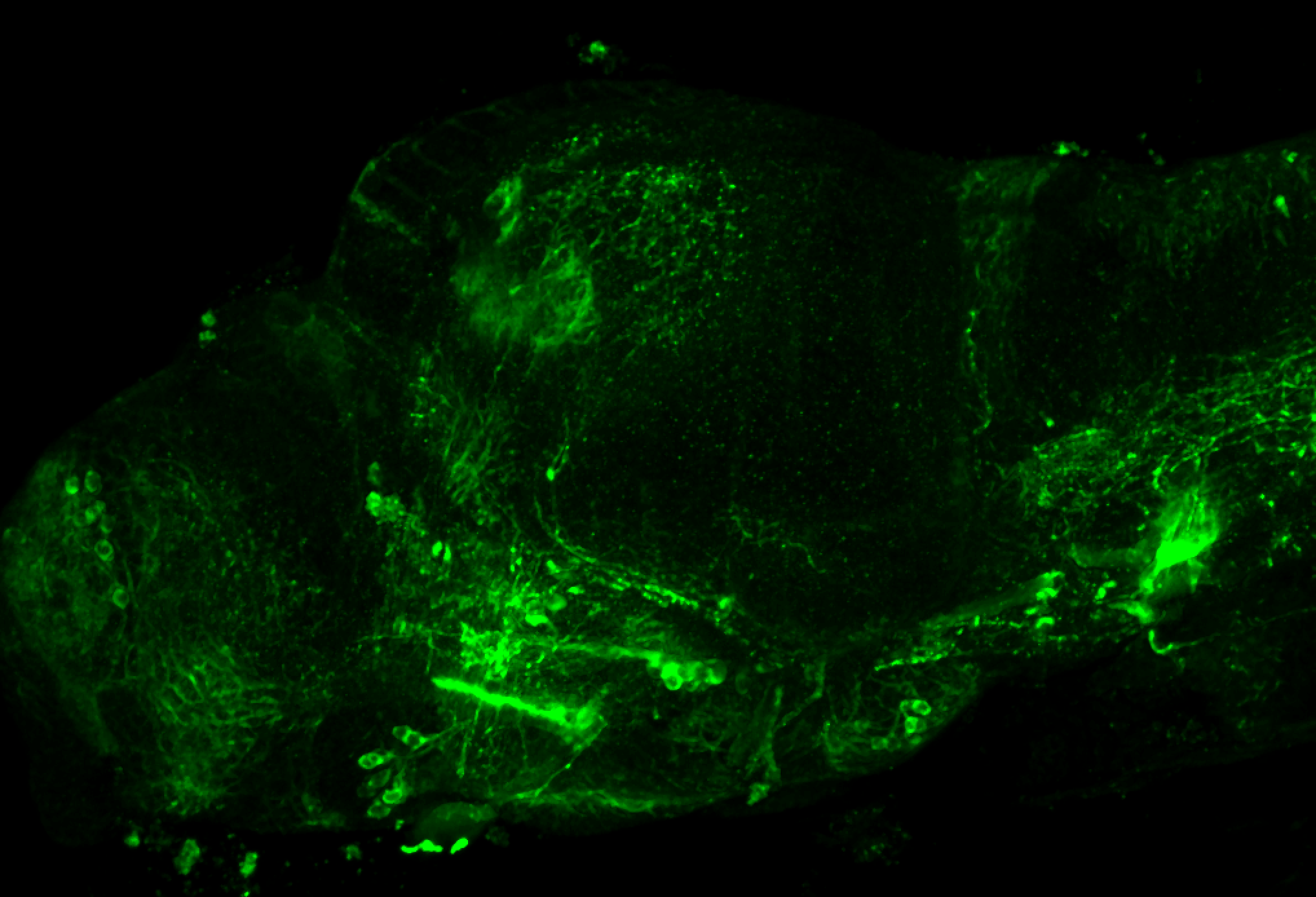
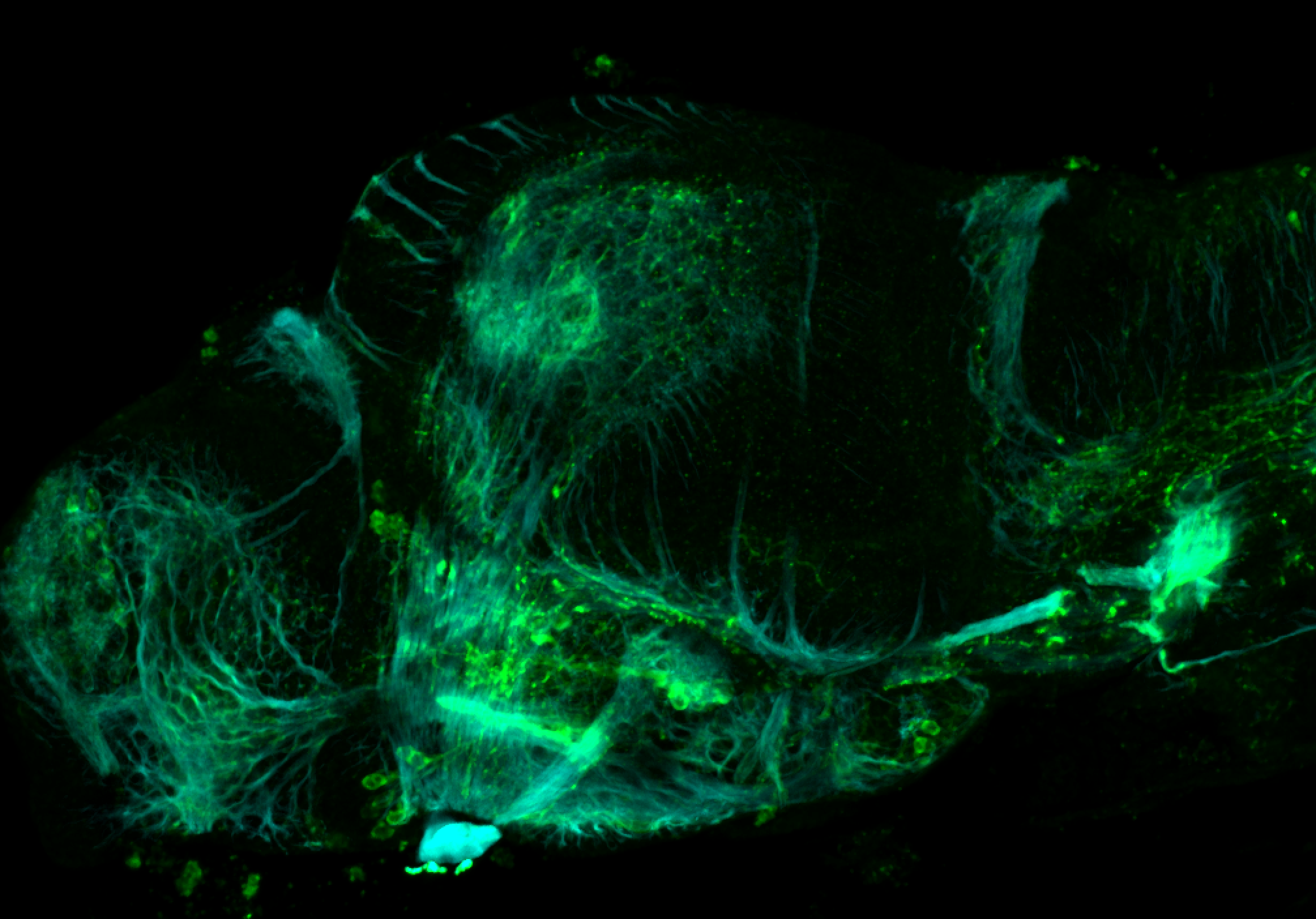
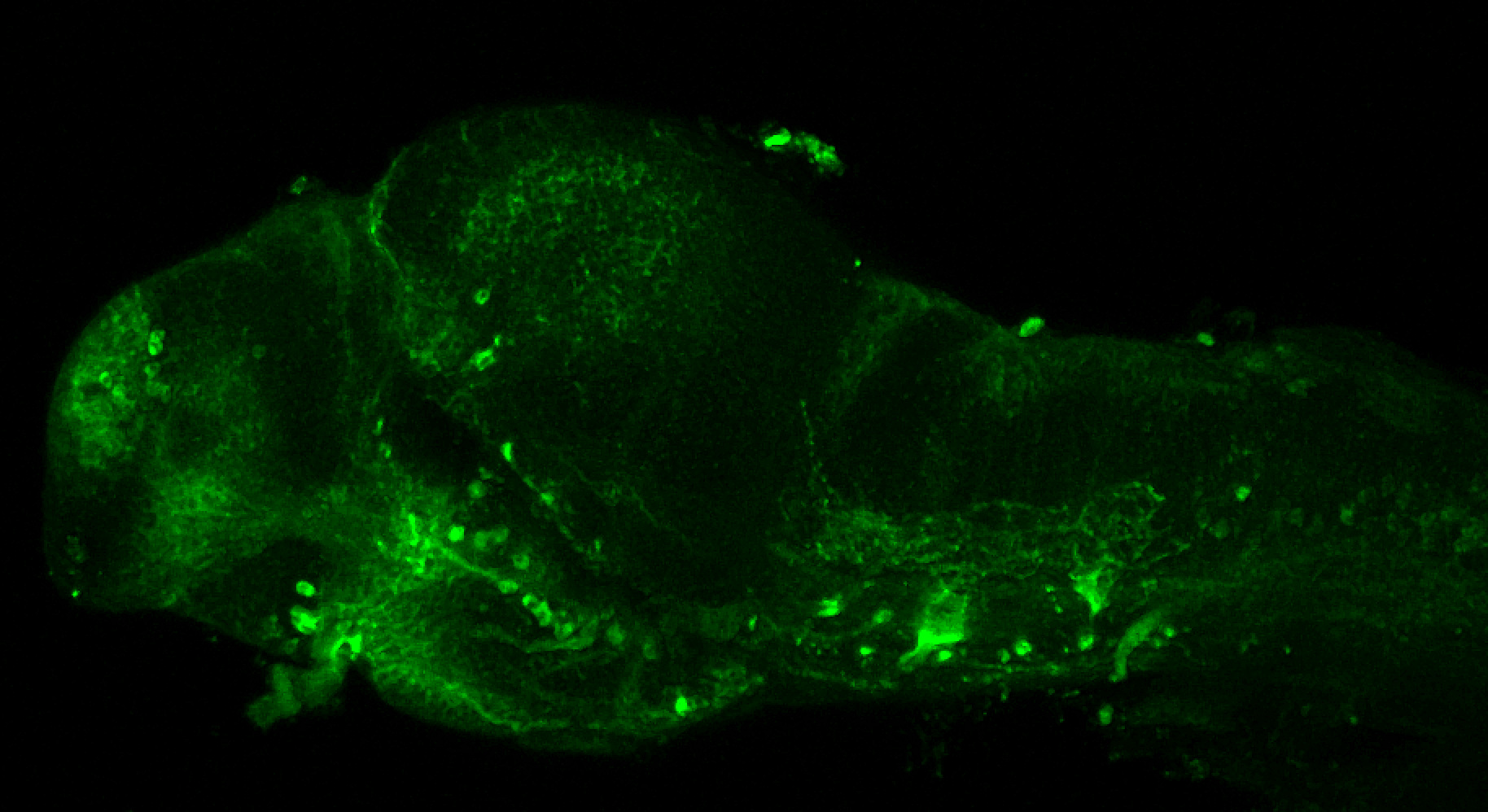
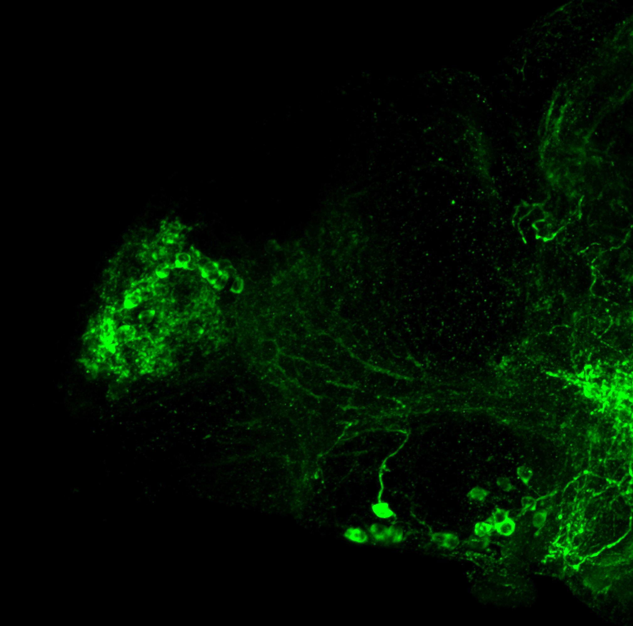
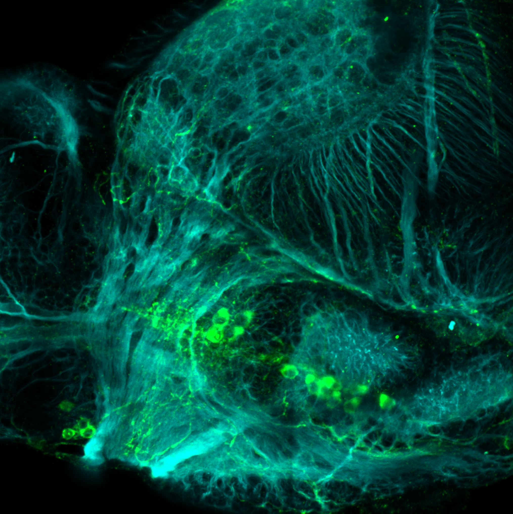
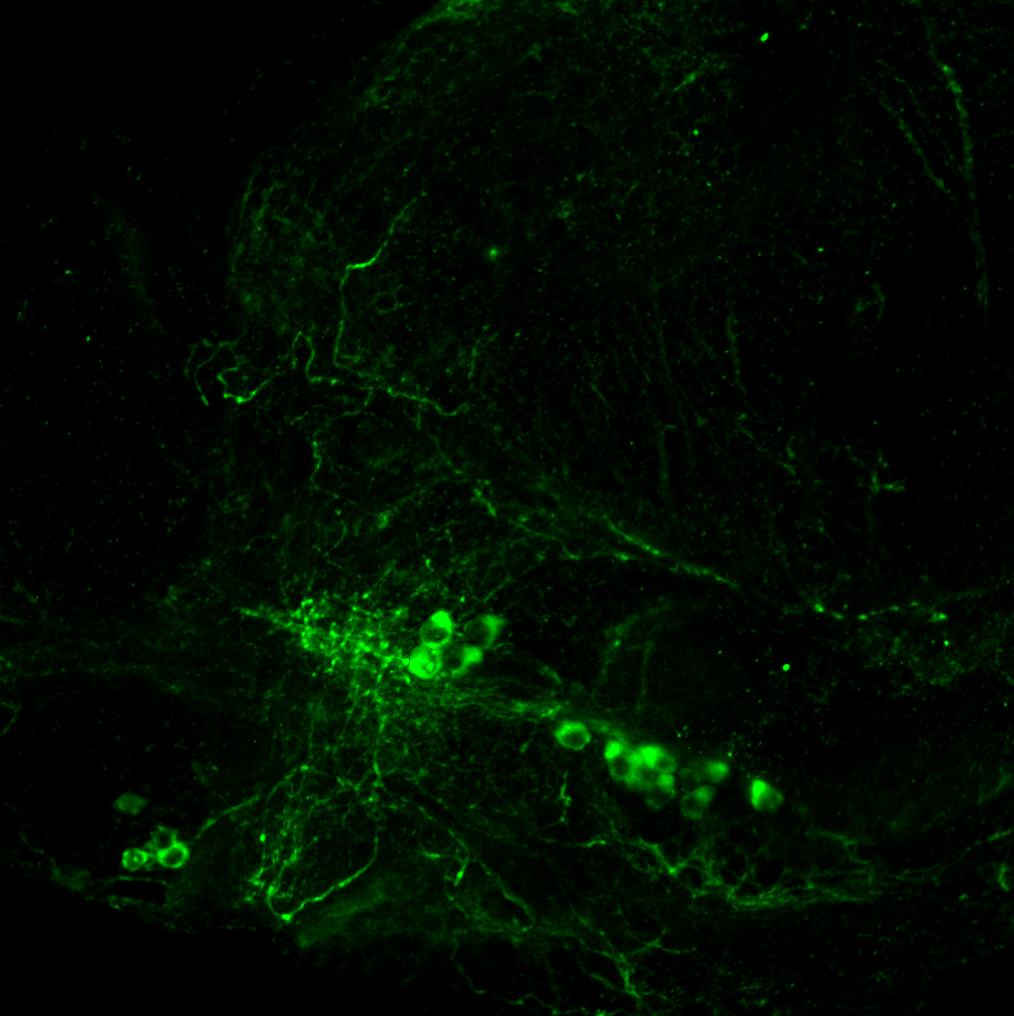
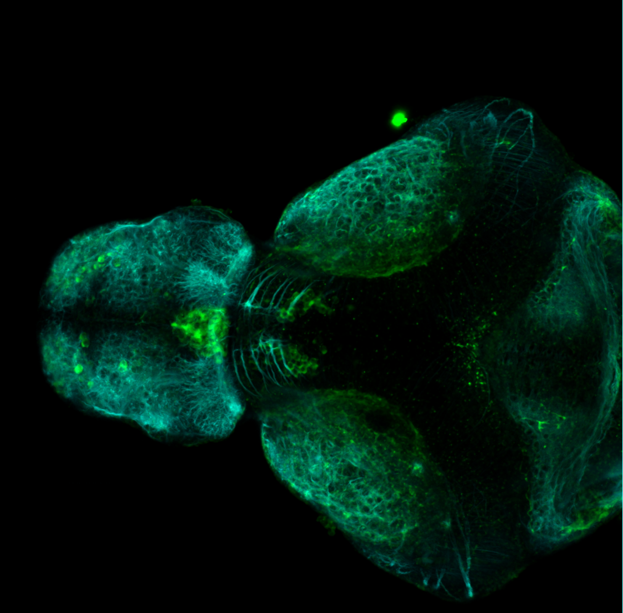
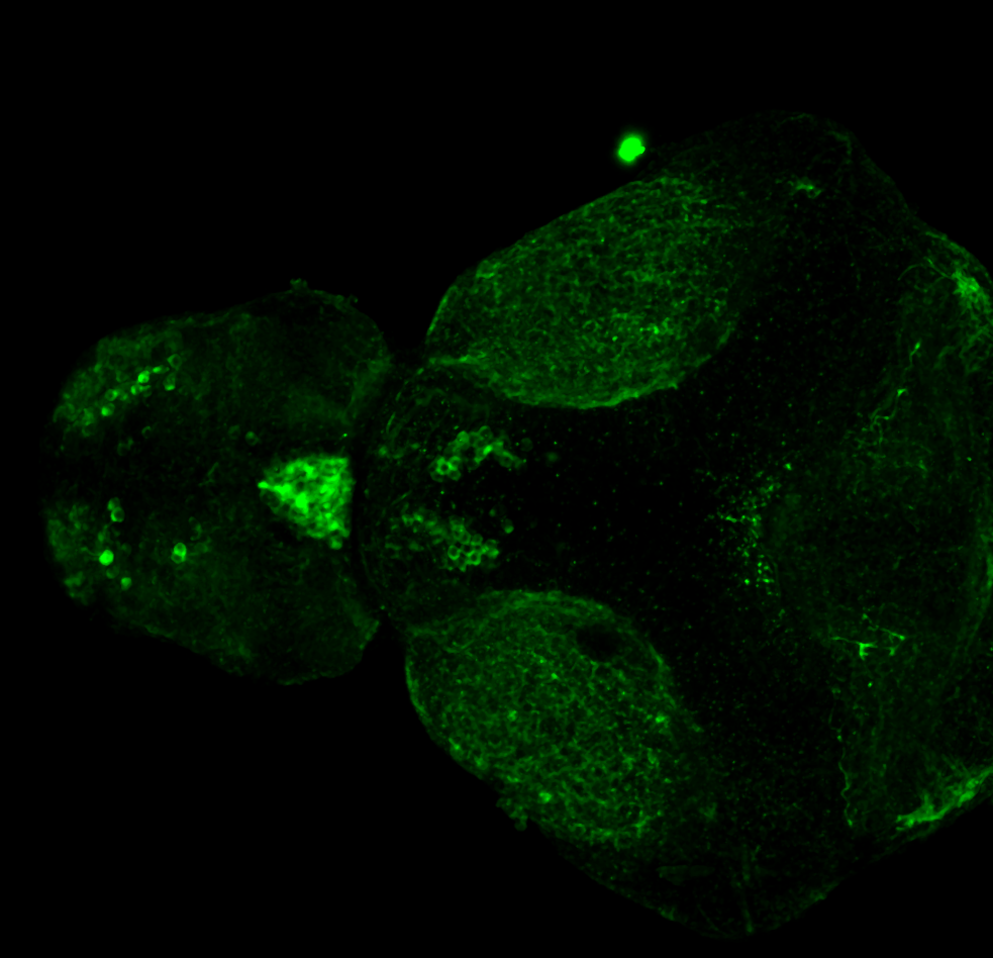
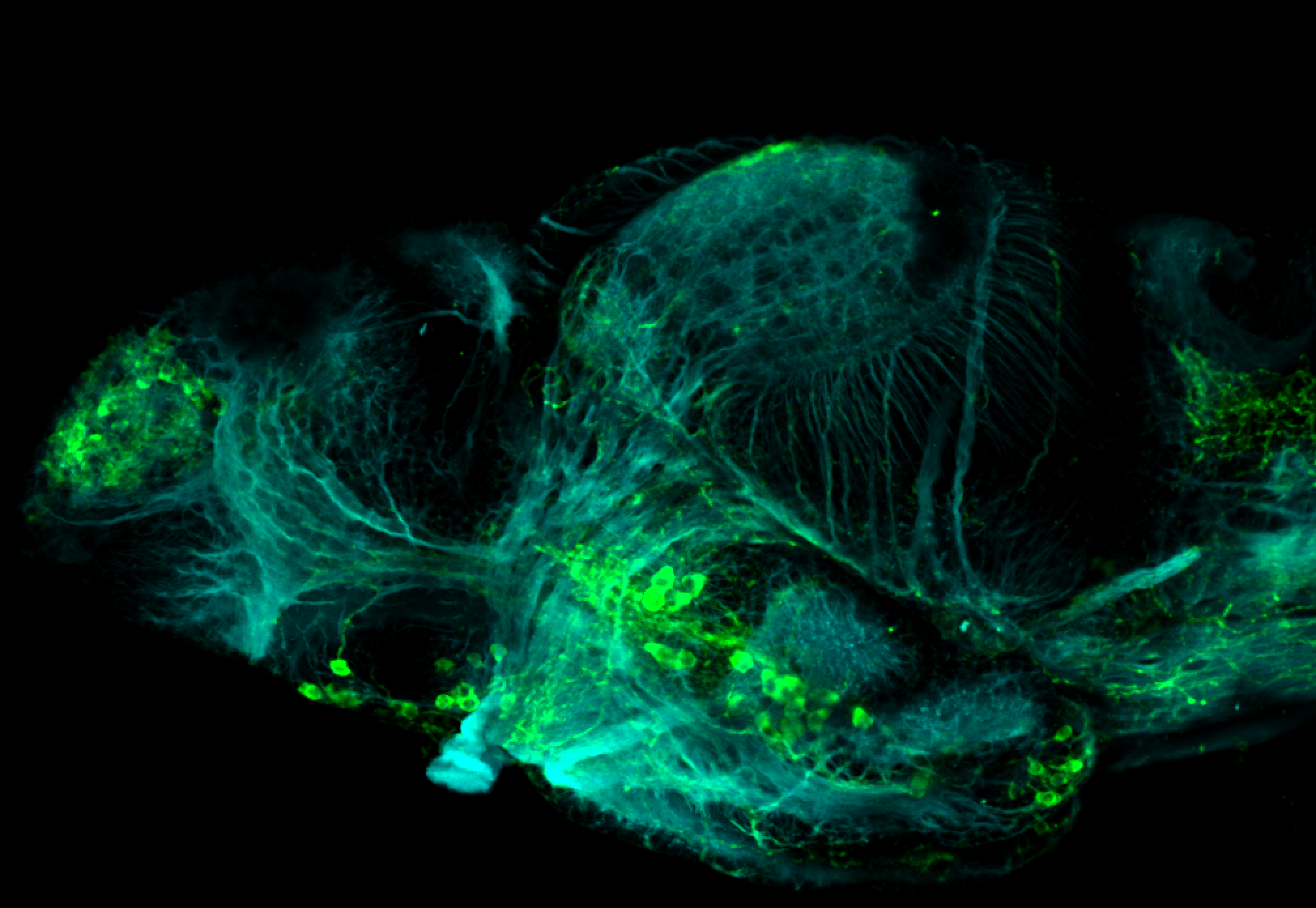
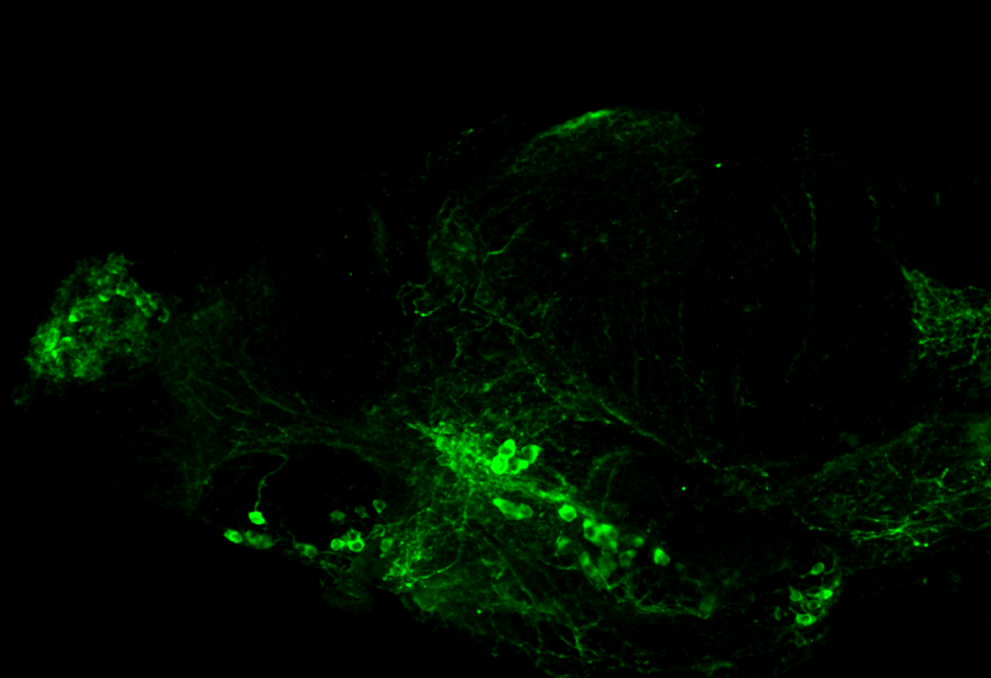
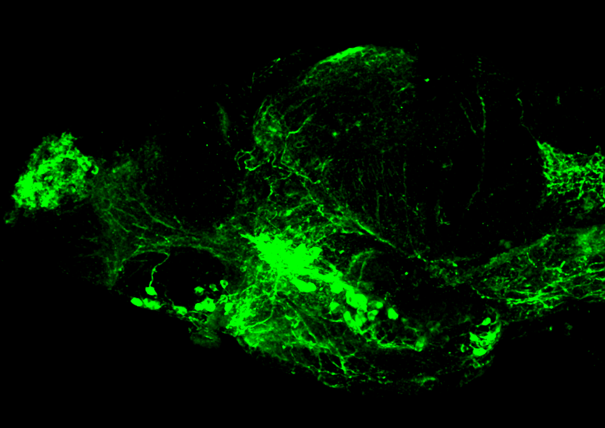
3A10 labels mauthner cells, reticulospinal neurons and lateral line in the hindbrain.

Mouse monoclonal anti-3A10 (IgG1) (DSHB, Cat#AB_531874, dilution 1:200)
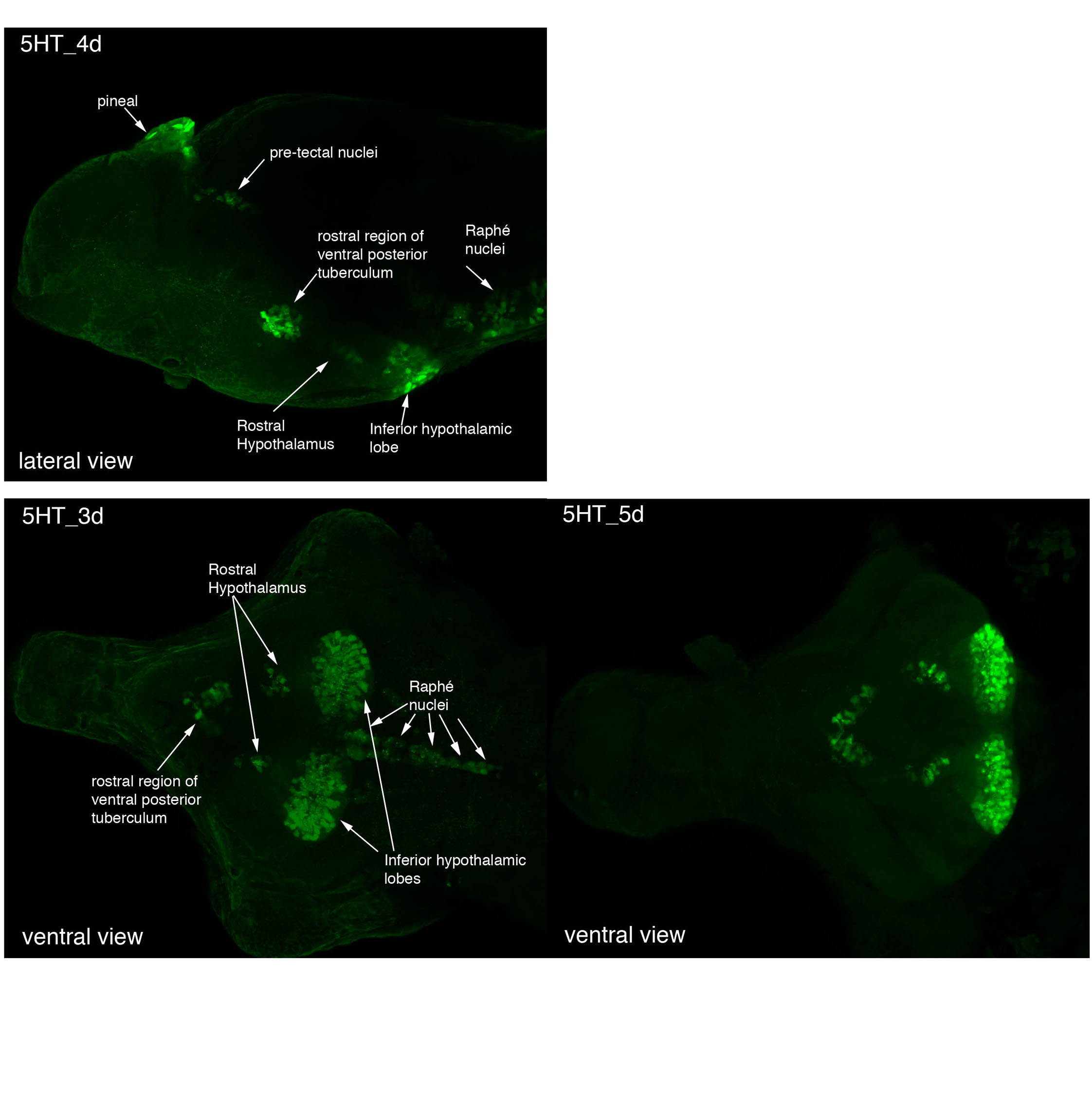
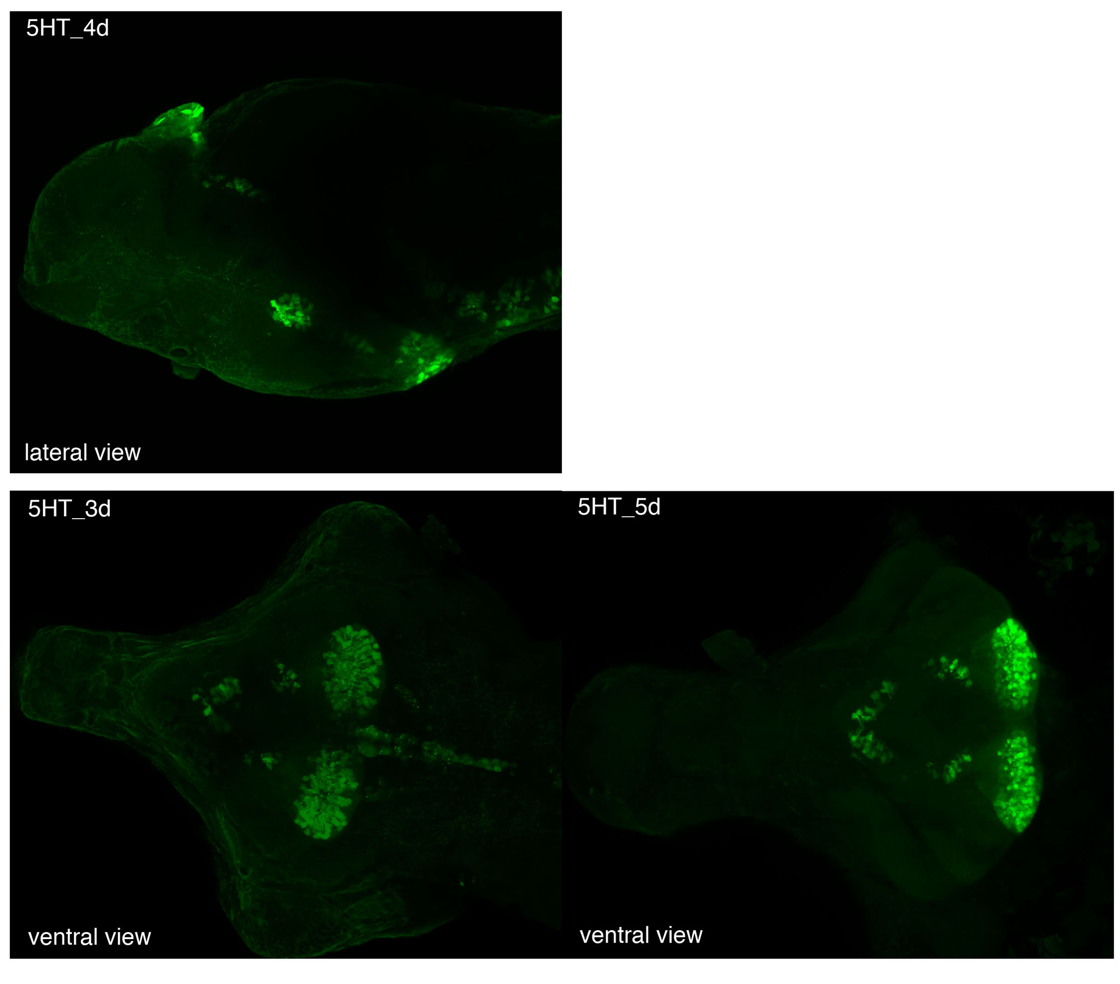
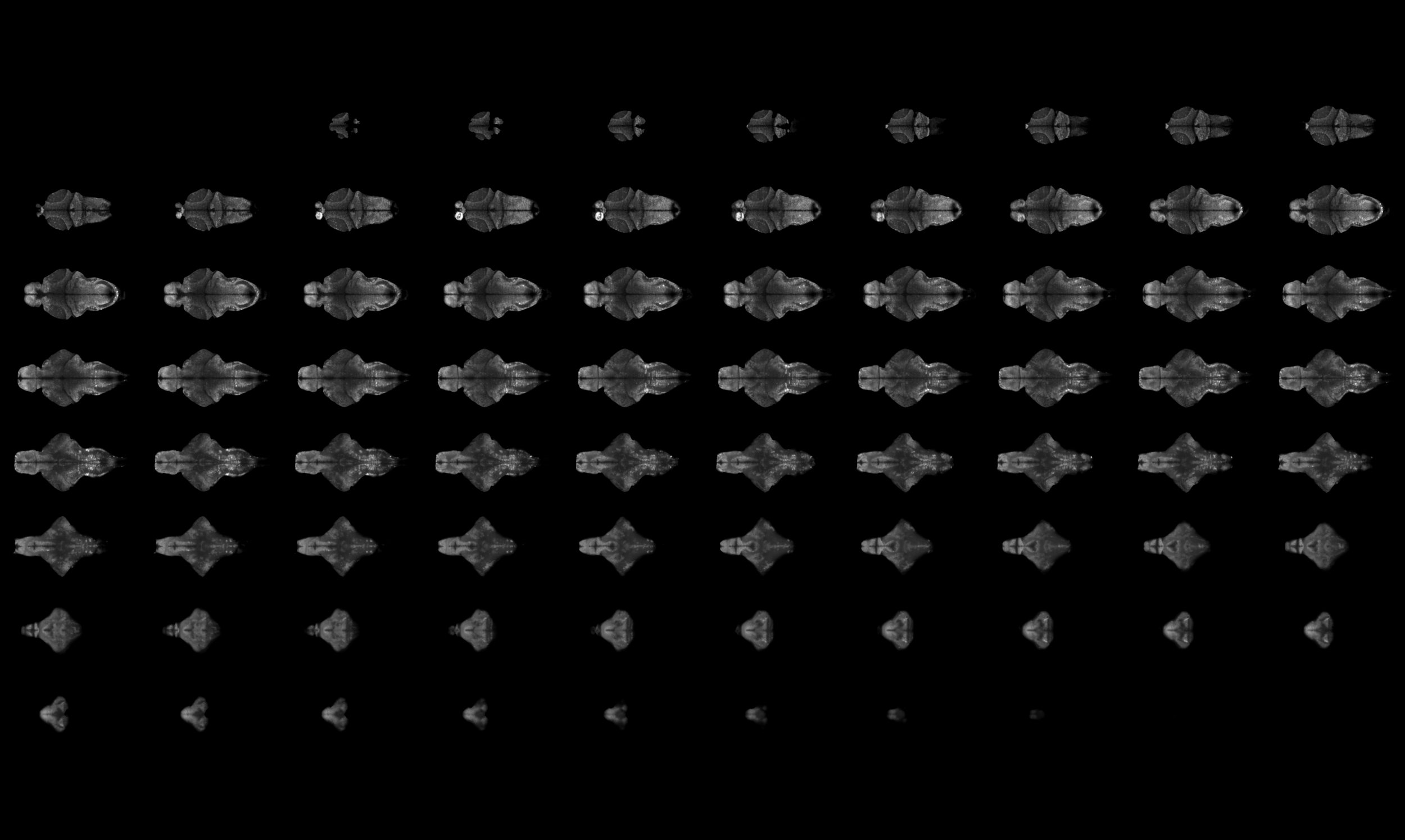
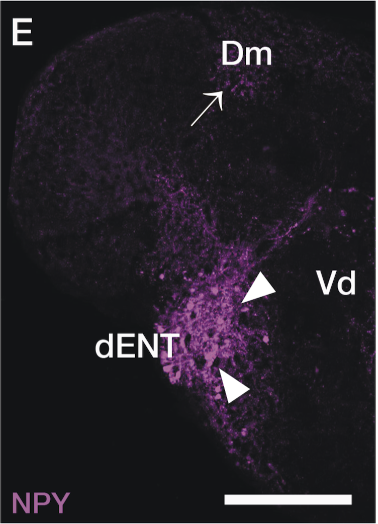
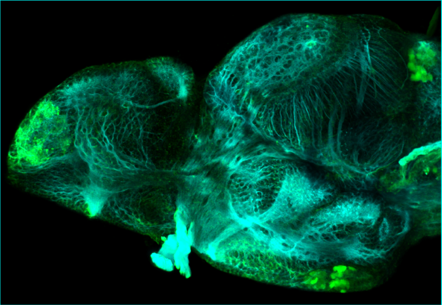
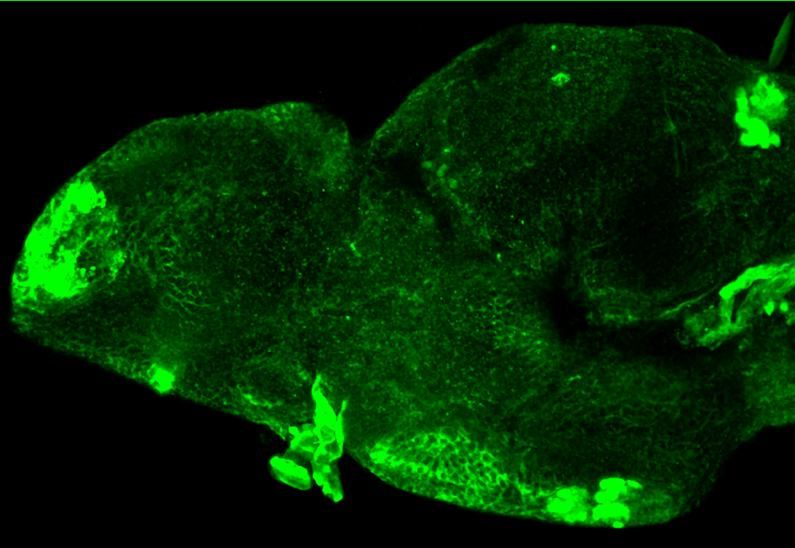
Select images

External Links:

 ZFIN


LABELS THESE BRAIN STRUCTURES:

mauthner neurons, reticulospinal neurons, lateral line.


KEY PUBLICATIONS

Brand, M., Heisenberg, C.P., Jiang, Y.J., Beuchle, D., Lun, K., Furutani-Seiki, M., Granato, M., Haffter, P., Hammerschmidt, M., Kane, D.A., Kelsh, R.N., Mullins, M.C., Odenthal, J., van Eeden, F.J., and Nüsslein-Volhard, C. (1996)
Mutations in zebrafish genes affecting the formation of the boundary between midbrain and hindbrain. Development (Cambridge, England). 123:179-190. 

5-HT (serotonin)

5-HT (serotonin)

5-HT(serotonin)

ABOUT THIS ANTIBODY

5-HT labels serotinergic neurons in the pineal organ, pretectum, posterior tuberculum, hypothalamus and superior and inferior raphe.

Rat monoclonal anti-5-HT (IgG) (Chemicon/Millipore, Cat#MAB352, dilution 1:500)

Select images

External Links:

 ZFIN


LABELS THESE BRAIN STRUCTURES:

 pineal organ, pretectum, posterior tuberculum, hypothalamus, superior raphe, inferior raphe.


KEY PUBLICATIONS

Lillesaar.
The serotonergic system in fish.
J Chem Neuroanat (2011) vol. 41 (4) pp. 294-308

Gaspar & Lillesaar.
Probing the diversity of serotonin neurons.
Philos Trans R Soc Lond B Biol Sci. 2012 Sep 5;367(1601):2382-94. doi: 10.1098/rstb.2011.0378.

Lillesaar et al.
Axonal projections originating from raphe serotonergic neurons in the developing and adult zebrafish, Danio rerio, using transgenics to visualize raphe-specific pet1 expression.
J Comp Neurol. 2009 Jan 10;512(2):158-82. doi: 10.1002/cne.21887.

Panula et al.
Modulatory neurotransmitter systems and behavior: towards zebrafish models of neurodegenerative diseases.
Zebrafish (2006) vol. 3 (2) pp. 235-47

McLean and Fetcho.
Ontogeny and innervation patterns of dopaminergic, noradrenergic, and serotonergic neurons in larval zebrafish.
J. Comp. Neurol. (2004) vol. 480 (1) pp. 38-56

Wen et al.
Visualization of monoaminergic neurons and neurotoxicity of MPTP in live transgenic zebrafish. Developmental Biology (2008) vol. 314 (1) pp. 84-92 

Acetylcholinesterase (AchE)

Acetylcholinesterase (AchE)

Acetylcholinesterase

ABOUT THIS LABELLING

AChE is believed to be present transiently in neurons, including non-cholinergic ones, soon after they complete their final mitosis, and therefore has been used as a marker of differentiation (Wilson et al., 1990).

METHOD FROM Wilson et al., 1990.

We used the method of Karnovsky and Roots (1964). 1-day embryos were fixed in 4% formalin buffered with either maleate (pH6.0, 0.1M) or Pipes (disodium salt - Sigma, pH6.95, O.lM-plus 2mM-EGTA [JV,/V,/V',A''-tetraacetic acid, Sigma], and lmM-MgSO4). Embryos prepared as whole mounts (n=18) were incubated for 2.5 h in maleate buffer (pH6.0) with 5mM-sodium citrate, 3mM-copper sulphate, 0.5mM-potassium ferricyanide and 0.5mgml~' substrate (acetylthiocholine iodide).

Select images

All produced by the boss: Steve Wilson.

External Links:

 ZFIN:


LABELS THESE BRAIN STRUCTURES:

differentiated neurons, cholinergic neurons.  


KEY PUBLICATIONS

 Wilson et al., Wilson, S.W., Ross, L.S., Parrett, T., and Easter, S.S. Jr. (1990)
The development of a simple scaffold of axon tracts in the brain of the embryonic zebrafish, Brachydanio rerio.
Development (Cambridge, England). 108:121-145.

KARNOVSKY, M. J. AND ROOTS, L. (1964).
A direct coloring thiocholine method for cholinesterases.
J. Histocliem. Cytochem. 12, 219-221.

acetylated tubulin

acetylated tubulin

anti-acetylated tubulin

ABOUT THIS ANTIBODY

anti-acetylated tubulin labels axonal tracts. Please see our separate white matter section for more information about individual tracts and commissures. 

This is a beautiful antibody and like anti-SV2 makes a nice general counterstain when looking at neuroanatomy in transgenic larvae. We also use this antibody extensively to look for any defects in brain development in mutant larvae. 

Mouse monoclonal anti-acetylated tubulin (IgG2b) (alpha-tubulin, Sigma, Cat# T7451, dilution 1:250)

Select images

woodshole_tubulin_labelled.gif
woodshole_tubulin2_labelled.gif

External Links:

 ZFIN:https://zfin.org/ZDB-ATB-081003-6


LABELS THESE BRAIN STRUCTURES:

Axonal tracts. 


KEY PUBLICATIONS

Wilson, S.W., Ross, L.S., Parrett, T., and Easter, S.S. Jr. (1990)
The development of a simple scaffold of axon tracts in the brain of the embryonic zebrafish, Brachydanio rerio. 
Development (Cambridge, England). 108:121-145

Chitnis, A.B. and Kuwada, J.Y. (1990) 
Axonogenesis in the brain of zebrafish embryos. 
The Journal of neuroscience : the official journal of the Society for Neuroscience. 10:1892-1905

Karlstrom, R.O., Trowe, T., Klostermann, S., Baier, H., Brand, M., Crawford, A.D., Grunewald, B., Haffter, P., Hoffman, H., Meyer, S.U., Muller, B.K., Richter, S., van Eeden, F.J., Nüsslein-Volhard, C., and Bonhoeffer, F. (1996) 
Zebrafish mutations affecting retinotectal axon pathfinding. 
Development (Cambridge, England). 123:427-438

Calbindin

Calbindin

Calbindin D-28k

ABOUT THIS ANTIBODY

anti-Calbindin recognises a calcium-binding protein of the EF-hand family related to calmodulin and troponin-C. This antibody labels the olfactory epithelia, trigeminal nerve, and cells in the optic tectum and pretectum.

Mouse monoclonal anti-Calbindin D-28k (IgG1) (Swant, Cat#CB300, dilution 1:200).

Select images

External Links: http://www.swant.com/pdfs/Monoclonal_anti_calbindin_d28k_300.pdf

 ZFIN


LABELS THESE BRAIN STRUCTURES:

 olfactory epithelium, olfactory bulb, eminentia thalami, RGC, optic nerve, pretectum, optic tectum, hypothalamus, cerebellum, horizontal commissure.


KEY PUBLICATIONS


Calretinin

Calretinin

Rabbit anti-calretinin

ABOUT THIS ANTIBODY

anti-calretinin labels neurons expressing the calcium binding protein calretinin.

Calcium-binding preoteins contribute to calcium homeostasis by buffering intercellular calcium. This antibody labels distinct groups of neurons in the central and peripheral nervous system.
 

Rabbit Polyclonal anti-calretinin (Swant, Cat#CR7697, dilution 1:1000)

SELECT IMAGES

EXTERNAL LINKS:

 ZFIN:https://zfin.org/ZDB-ATB-160726-1
Swant: https://www.swant.com/pdfs/Rabbit_anti_calretinin_7697.pdf


LABELS THESE BRAIN STRUCTURES:

olfactory epithelium, olfactory glomeruli, dorsal telencephalon, ventral telencephalon, entopeduncular nucleus, preoptic area, preglomerular complex, mammillary body, hypothalamus (inferior lobe), torus lateralis, posterior tuberculum, thalamus(anterior thalamic nucleus), pretectum (parvocellular superficial pretectal nucleus), nucleus of the medial longitudinal fascicle. 


KEY PUBLICATIONS

Castro A1, Becerra M, Manso MJ, Anadón R.
Calretinin immunoreactivity in the brain of the zebrafish, Danio rerio: distribution and comparison with some neuropeptides and neurotransmitter-synthesizing enzymes. I. Olfactory organ and forebrain.
J Comp Neurol. 2006 Jan 20;494(3):435-59.

Beretta, C.A., Dross, N., Guglielmi, L., Bankhead, P., Soulika, M., Gutierrez-Triana, J.A., Paolini, A., Poggi, L., Falk, J., Ryu, S., Kapsimali, M., Engel, U., Carl, M. (2017)
Early Commissural Diencephalic Neurons Control Habenular Axon Extension and Targeting.
Current biology : CB. 27(2):270–278.

Turner, K.J., Hawkins, T.A., Yáñez, J., Anadón, R., Wilson, S.W., Folgueira, M. (2016)
Afferent Connectivity of the Zebrafish Habenulae.
Frontiers in neural circuits 10:30. https://doi.org/10.3389/fncir.2016.00030

GABA (gamma-Aminobutyric acid)

GABA (gamma-Aminobutyric acid)

GABA

ABOUT THIS ANTIBODY

anti-GABA labels GABA-ergic neurons. Gamma-Aminobutyric acid is an inhibitory neurotransmitter. GABA-ergic synaptic transmission reduces neuronal excitability in the post-synatic neuron by hyperpolarising the cell through the opening of GABA receptor mediated ion channels which allows chloride ions to flow into the or potassium ions to flow out of the postsynaptic neuron. .

Rabbit Polyclonal anti-GABA (Sigma, Cat#A2052, dilution 1:1000)

NB: this antibody requires fixation to be very short (Max of 15 mins for dissected embryos).

Select images

External Links:

 ZFIN


LABELS THESE BRAIN STRUCTURES:

 gabaergic neurons,


KEY PUBLICATIONS

Mueller, T., Vernier, P., and Wullimann, M.F. (2006)
A phylotypic stage in vertebrate brain development: GABA cell patterns in zebrafish compared with mouse.
The Journal of comparative neurology. 494(4):620-634

HuC/D

HuC/D

HuC/D

ABOUT THIS ANTIBODY

HuC/D is a pan-neuronal nuclear label and as such a very good counterstain for showing the structure of the brain.


anti-HuC/D (Manufacturer/cat#/dilution)

Select images by Chintan Trivedi imaged using a light-sheet microscope.

Flythrough movie of a 6dpf larval zebrafish brain labelled with anti-HuC and anti-HuD antibodies. Anti-HuC/D is a pan-neuronal nuclear label that labels all neurons in the nervous system.

Flythrough movie of a 6dpf larval zebrafish brain labelled with anti-HuC and anti-HuD antibodies. Anti-HuC/D is a pan-neuronal nuclear label that labels all neurons in the nervous system.


Montage of slices through the brain of a 6dpf zebrafish larvae labelled using anti-HuC/D antibody.

Montage of slices through the brain of a 6dpf zebrafish larvae labelled using anti-HuC/D antibody.


6dpf brain of a larval zebrafish labeled with anti-HuC/D. This stack was morphed to ZBB. This screenshot shows the HuC/D antibody labelling uploaded to ZBB and viewed using their online browser. If you would like to look at this data using ZBB pleas…

6dpf brain of a larval zebrafish labeled with anti-HuC/D. This stack was morphed to ZBB. This screenshot shows the HuC/D antibody labelling uploaded to ZBB and viewed using their online browser. If you would like to look at this data using ZBB please download the .png file using the link below and upload it to ZBB using the Lines> Custom>Load option.

External Links:

 ZFIN:

Download the HuC/D .png file to view the data using Zebrafish Brain Browser


LABELS THESE BRAIN STRUCTURES:

 pan-neuronal marker.


KEY PUBLICATIONS

NPY (neuropeptide Y)

NPY (neuropeptide Y)

NPY

ABOUT THIS ANTIBODY

NPY labels neurons that use neuropeptide Y as a neurotransmitter. This antibody labels the dorsal entopeduncular nucleus.

Rabbit anti-NPY (Sigma, Cat#N9528, dilution 1:1000)

Select images

External Links:

 ZFIN:


LABELS THESE BRAIN STRUCTURES:

 dorsal entopeduncular nucleus,


KEY PUBLICATIONS

Turner, K.J., Hawkins, T.A., Yáñez, J., Anadón, R., Wilson, S.W., Folgueira, M. (2016)
Afferent Connectivity of the Zebrafish Habenulae.
Frontiers in neural circuits. 10:30

Singh, C., Rihel, J., Prober, D.A. (2017)
Neuropeptide Y Regulates Sleep by Modulating Noradrenergic Signaling.
Current biology : CB. 27(24):3796-3811.e5.

Parvalbumin

Parvalbumin

Parvalbumin

ABOUT THIS ANTIBODY

anti-Parvalbumin labels .

Parvalbumin is a calcium-binding albumin protein found in brain and muscle. This antibody has relatively sparse labelling in the larval zebrafish brain. It nicely labels the olfactory epithelium, olfactory sensory nerve processes and the olfactory nerves, neuromasts of the lateral line system, amacrine cells in the retina and a subset of purkinje cells in the cerebellum and the pituitary. Parvalbumin also labels neurons in the optic tectum at adult stages (Robra et al., 2016).

Mouse monoclonal anti-Parvalbumin (IgG1) (Chemicon/Millipore, Cat#MAB1572, dilution 1:200)

Select images

All images by Monica Folgueira

External Links:

 ZFIN


LABELS THESE BRAIN STRUCTURES:

olfactory epithelium, olfactory nerve, olfctory bulb, amacrine cell in retina, pituitary, cerebellum, purkinje cell, optic tectum.  


KEY PUBLICATIONS

 
Yeo, J.Y., Lee, E.S., and Jeon, C.J. (2009)
Parvalbumin-immunoreactive neurons in the inner nuclear layer of zebrafish retina.
Experimental Eye Research. 88(3):553-560.

Kaslin, J., Kroehne, V., Benato, F., Argenton, F., and Brand, M. (2013)
Development and specification of cerebellar stem and progenitor cells in zebrafish: from embryo to adult. Neural Development. 8(1):9.

Kress, S., Biechl, D., Wullimann, M.F. (2015)
Combinatorial analysis of calcium-binding proteins in larval and adult zebrafish primary olfactory system identifies differential olfactory bulb glomerular projection fields.
Brain structure & function. 220(4):1951-70.

synaptic vesicle 2 (SV2)

synaptic vesicle 2 (SV2)

Synaptic Vesicle 2 (SV2)

ABOUT THIS ANTIBODY

Synaptic Vesicle 2 labels synaptic neuropil.

We frequently use this antibody in conjunction with  anti-acetylated tubulin (IgG2b, Sigma), which labels beautifully the axonal connections, to aid anatomical orientation in transgenic specimens. In addition to these antibodies being informative from a neuroanatomical perspective, they are also invaluable as a tool for anatomical localisation. These antibodies can be used as a framework to easily compare the expression patterns of different transgenic lines and locate GFP-positive structure in the context of the brain. Both antibodies are mouse monoclonals but can be detected in the same specimen using subtype-specific secondary antibodies.

Mouse monoclonal anti-SV2 (IgG1) (DSHB, Cat#AB 2315387, dilution 1:500)

Select images

External Links:

 ZFIN:https://zfin.org/ZDB-ATB-081201-1


LABELS THESE BRAIN STRUCTURES:

Synaptic neuropil


KEY PUBLICATIONS

Buckley K, Kelly RB (1985)
Identification of a transmembrane glycoprotein specific for secretory vesicles of neural and endocrine cells. 
J Cell Biol 100:1284–1294

Hendricks M, Jesuthasan S (2007)
Asymmetric innervation of the habenula in zebrafish. 
J Comp Neurol 502:611–619

Turner KJ1, Bracewell TG, Hawkins TA.
Anatomical dissection of zebrafish brain development.
Methods Mol Biol. 2014;1082:197-214. doi: 10.1007/978-1-62703-655-9_14.

Tyrosine Hydroxylase

Tyrosine Hydroxylase

Tyrosine Hydroxylase

ABOUT THIS ANTIBODY

Tyrosine Hydroxylase is an enzyme that catalyses the convertion of L-tyrosine to L-3,4-dihydroxyphenylalanine(L-DOPA) which is the precursor for the neurotransmitter dopamine. anti-TH labels dopaminergic neurons in the olfactory bulb, subpallium, preotic area, posterior tuberculum and locus coeruleus.

Rabbit polyclonal anti-TH (Chemicon/Millipore , Cat#AB152, dilution 1:1000)

Select images

External Links:

 ZFIN


LABELS THESE BRAIN STRUCTURES:

 olfactory bulb, preoptic area, posterior tuberculum.


KEY PUBLICATIONS

 Yamamoto, K., Ruuskanen, J.O., Wullimann, M.F., and Vernier, P. (2010)
Two tyrosine hydroxylase genes in vertebrates: New dopaminergic territories revealed in the zebrafish brain. Molecular and Cellular Neuroscience. 43(4):394-402.

Yamamoto, K., Ruuskanen, J.O., Wullimann, M.F., and Vernier, P. (2011)
Differential expression of dopaminergic cell markers in the adult zebrafish forebrain.
The Journal of comparative neurology. 519(3):576-598.

Xi, Y., Yu, M., Godoy, R., Hatch, G., Poitras, L., and Ekker, M. (2011)
Transgenic zebrafish expressing green fluorescent protein in dopaminergic neurons of the ventral diencephalon.
Developmental dynamics. 240(11):2539-47.